Article

Rapid Decline of Serum Proprotein Convertase Subtilisin/Kexin 9 (PCSK9) in Non-Cirrhotic Patients with Chronic Hepatitis C Infection Receiving Direct-Acting Antiviral Therapy

1
Department of Internal Medicine I, Gastroenterology, Hepatology, Endocrinology, Rheumatology and Infectious Diseases, University Hospital Regensburg, 93053 Regensburg, Germany
2
Department of Visceral Surgery and Medicine, University Inselspital, 3010 Bern, Switzerland
*
Author to whom correspondence should be addressed.
Submission received: 22 February 2021 / Revised: 30 March 2021 / Accepted: 6 April 2021 / Published: 11 April 2021
(This article belongs to the Section Gastroenterology & Hepatopancreatobiliary Medicine)

Abstract

:
Direct-acting antivirals (DAAs) efficiently eradicate the hepatitis C virus (HCV). Low-density lipoprotein (LDL) levels increase rapidly upon DAA treatment. Proprotein convertase subtilisin/kexin 9 (PCSK9) induces degradation of the hepatic LDL receptor and thereby elevates serum LDL. The aim of this study was to determine serum PCSK9 concentrations during and after DAA therapy to identify associations with LDL levels. Serum PCSK9 was increased in 82 chronic HCV-infected patients compared to 55 patients not infected with HCV. Serum PCSK9 was low in HCV patients with liver cirrhosis, but patients with HCV-induced liver cirrhosis still exhibited higher serum PCSK9 than patients with non-viral liver cirrhosis. Serum PCSK9 correlated with measures of liver injury and inflammation in cirrhotic HCV patients. In patients without liver cirrhosis, a positive association of serum PCSK9 with viral load existed. Serum PCSK9 was not different between viral genotypes. Serum PCSK9 did not correlate with LDL levels in HCV patients irrespective of cirrhotic status. Serum PCSK9 was reduced, and LDL was increased at four weeks after DAA therapy start in non-cirrhotic HCV patients. Serum PCSK9 and LDL did not change upon DAA treatment in the cirrhotic group. The rapid decline of PCSK9 after the start of DAA therapy in conjunction with raised LDL levels in non-cirrhotic HCV patients shows that these changes are not functionally related.

Graphical Abstract

1. Introduction

Chronic hepatitis C virus (HCV) infection is a risk factor for liver fibrosis, cirrhosis and hepatocellular carcinoma [1]. HCV is successfully eliminated by the use of direct-acting antivirals (DAA), and sustained virologic response (SVR) of up to 100% can be achieved [2]. HCV eradication normalizes serum levels of aminotransferases and ferritin, whereas bilirubin and albumin levels are barely changed [3,4,5,6,7]. DAA therapy results in short-term improvements of hepatic inflammation but not fibrosis [8].
Of note, HCV reprograms the host lipoprotein metabolism for the production of viral particles. Indeed, expression of the LDL-receptor was increased in HCV-infected livers [9,10]. Accordingly, serum cholesterol and low-density lipoprotein (LDL) levels were low in patients with chronic HCV infection [11,12]. Upon DAA therapy, serum LDL rapidly recovers [5,13], showing that lipid metabolism’s perturbation is a direct effect of HCV infection.
Proprotein convertase subtilisin/kexin 9 (PCSK9) is an enzyme predominantly produced by the liver [14]. Hepatic LDL-receptor protein is degraded upon PCSK9 binding, and serum LDL is increased. Blockage of PCSK9 lowers serum LDL and may protect from cardiovascular diseases [15,16,17].
HCV uses the LDL-receptor to enter hepatocytes [18,19]. PCSK9 participates in the degradation of the LDL-receptor and probably has a role in HCV infection [18,19]. Indeed, an inhibitory function of PCSK9 on HCV infection has been described [18,20]. Alirocumab, a therapeutic human anti-PCSK9 antibody, was, however, therapeutically ineffective [21].
Of clinical relevance, HCV-infected patients had higher serum PCSK9 levels, whereas LDL did not increase in parallel [22]. Serum PCSK9 negatively correlated with the model for end-stage liver disease (MELD) score, which is the state-of-the-art scoring model of liver disease severity [22,23]. In addition, HCV genotype-dependent regulation of plasma PCSK9 levels has been described. PCSK9 concentrations were lower in HCV genotype 3 patients compared to HCV-negative controls. HCV genotype 1-infected patients had higher PCSK9 levels than non-infected controls [24]. PCSK9 did not correlate with LDL in HCV genotype 1- or 3-infected patients [24].
LDL levels rapidly increase during DAA treatment [5,13,25]. Whether PCSK9 is induced in parallel is still poorly understood.
The studies cited above measured total PCSK9 protein by ELISA. PCSK9 is inactivated by proteolysis, and up to 40% of circulating PCSK9 is inactive [26]. One study reported that active PCSK9 declined, and the inactive form increased at week four after the start of daclatasvir/asunaprevir therapy in patients infected with HCV genotype 1b. At 24 weeks after the start of therapy, active and inactive PCSK9 concentrations returned to pretreatment levels. At 28 weeks posttreatment, active PCSK9 was higher compared to levels before therapy starts. Inactive PCSK9 was not changed during this period. In the serum of HCV patients, levels of inactive PCSK9 were about five-fold lower than the concentration of the active isoform [27]. Thus, total PCSK9 declined at four weeks after therapy start and steadily increased afterward. LDL was induced from week four after the start of therapy to six months posttreatment. These authors suggested that active PCSK9 has a role in the recovery of LDL. Active PCSK9 and LDL levels did not correlate before therapy and at 52 weeks posttreatment [27].
The association of serum PCSK9 with HCV infection and HCV-related dyslipidemia is not completely understood. Here, serum PCSK9 was measured in controls and patients with chronic HCV infection and the effect of HCV eradication on serum PCSK9 was determined.

2. Materials and Methods

2.1. Study Cohort

This study was conducted at the University Hospital of Regensburg from October 2014 to September 2019. Patients older than 18 years with chronic hepatitis C participated in the study. Treatment naive patients eligible for DAA therapy based on the EASL guidelines were included [2]. Patients were excluded if they were coinfected with HBV or HIV.
Statin taking was stopped during therapy. Most of the patients were treated with DAAs early after approval of these therapies, and at that time, the drug–drug interaction of statins and DAAs was suggested to be higher than known today [28].
Cirrhosis diagnosis by ultrasound was based on coarse liver parenchyma, nodular liver surface, and small liver size [29].
The fasting serum of the patients was collected in the morning. Serum aliquots were stored at −80 °C. Routine laboratory parameters were determined at the Institute of Clinical Chemistry and Laboratory Medicine (University Hospital Regensburg). All patients had chronic hepatitis C and were treated with one of the following regimens: sofosbuvir/daclatasvir, sofosbuvir/ledipasvir, glecaprevir/pibrentasvir, elbasvir/grazoprevir or sofosbuvir/velpatasvir, following international treatment guidelines [2].
The controls included patients referred to ultrasound imaging. Patients with cancers, severe liver diseases or liver cirrhosis were excluded. Data on PCSK9 serum levels in this control cohort were published before and are retrieved in this analysis for comparison with HCV-infected patients [10]. Moreover, analysis of PCSK9 in patients with predominant alcoholic liver cirrhosis was performed before, and data of patients with the non-viral disease were reused in this manuscript [10].
Patients’ characteristics are given in Table 1 and Table 2. All patients gave informed consent prior to inclusion in the study.

2.2. PCSK9 ELISA

The human PCSK9 DuoSet ELISA was obtained from R&D Systems (Wiesbaden, Nordenstadt, Germany) and was performed as requested by the provider (dilution of serum was 1:100). The serum used for analysis was thawed twice. The inter-assay coefficient of variation, which was determined from three independent experiments, was 3.4% for high values (serum of 8 patients diluted 1:10), 3.5% for median values (serum of 8 patients diluted 1:50) and 6.1% for low values (serum of 8 patients diluted 1:200). The intra-assay coefficient of variation was 4.3% for high values, 4.3% for median values and 7.6% for low values (Supplementary Materials). When serum of three donors was spiked with 1280 pg/mL recombinant PCSK9, recovery was 83%.

2.3. Statistical Analysis

Data are shown as boxplots. Small circles or asterisks above or below the boxes mark outliers. Statistical tests used were one-way ANOVA with post hoc Bonferroni, Mann–Whitney U-test, Spearman correlation, partial correlation (SPSS Statistics 25.0 program, IBM, Armonk, NY, USA) and t-test (Microsoft Excel). Two variables were compared by the chi-squared test. A value of p < 0.05 was regarded as significant.

3. Results

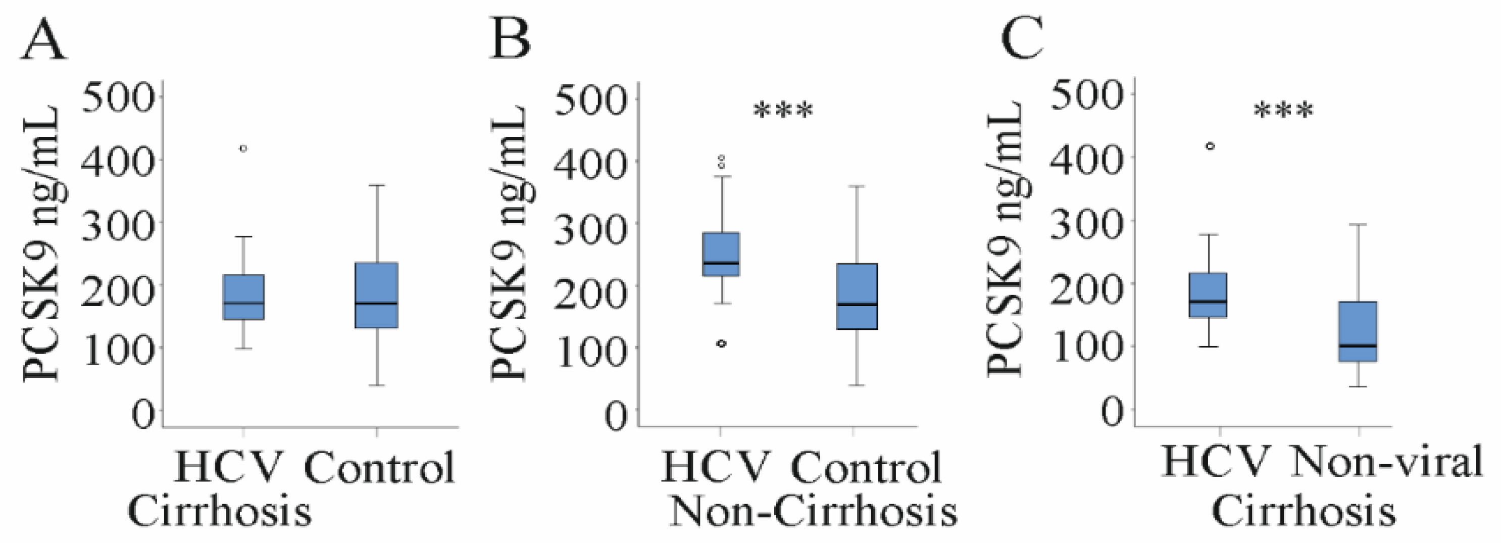
3.1. Serum PCSK9 Is Increased in HCV Patients

Serum PCSK9 was determined in 82 patients with chronic HCV infection. These samples were used recently for analysis of chemerin [5].
Comparison of serum PCSK9 between HCV patients and non-infected patients without any severe liver diseases showed that PCSK9 levels were 170 (40–360) ng/mL in the non-infected controls and 218 (99–418) ng/mL in HCV patients and were significantly higher in the HCV group (Figure 1A). Serum LDL was low in HCV patients compared to the control cohort as was described before [30] (Table 1).

3.2. Serum PCSK9 Correlates with Markers of Liver Injury, Viral Load and LDL in HCV Patients

Serum PCSK9 was similar in male and female HCV patients (Figure 1B) and did not correlate with body mass index (BMI) (Table 3). A modest negative association with age existed (Table 3). The 18 diabetic HCV patients had similar PCSK9 levels compared to the non-diabetic patients (Figure 1C).
Serum PCSK9 negatively correlated with bilirubin and INR, and accordingly with the MELD score (which is calculated from bilirubin, INR and creatinine [23]). There were modest negative associations with AST and CRP. Positive correlations existed for leukocyte count and for LDL (Table 3).
Serum PCSK9 was positively associated with viral load before therapy start (Table 3). HCV genotypes were 1a, 1b and 3a in 24, 38 and 14 patients, respectively. Six patients had different genotypes and were assigned to one group. Serum PCSK9 was similar in all of these patients (Figure 1D).
It must be noted that the prevalence of ultrasound diagnosed liver cirrhosis was similar between the different HCV genotypes and was 50% in patients with genotype 1a, 53% in patients with genotype 1b, 36% in patients with genotype 3a and 0% in patients with rare genotypes.

3.3. Serum PCSK9 in Relation to Liver Steatosis, Cirrhosis and Fibrosis Scores

Thirty-seven HCV patients had liver steatosis as was diagnosed by ultrasound imaging. Serum PCSK9 levels were not changed in patients having the fatty liver disease (Figure 2A). Non-invasive scores assessed liver fibrosis: (1) acoustic radiation force impulse (ARFI) indicated liver fibrosis in 67 patients, (2) the NAFLD score (which integrates age, BMI, diabetes, AST/ALT ratio, platelet count and albumin [31]) in 35 patients, (3) the aspartate aminotransferase/platelet (AST/PLT) ratio index (APRI) in 57 patients, and (4) the fibrosis 4 (FIB-4) score (calculated from age, AST, ALT and platelet count [32,33] in 53 patients (Table 1).
Increasing ARFI fibrosis score did not affect serum PCSK9 levels (Figure 2B). PCSK9 levels negatively correlated with fibrosis assessed by the FIB-4 score, the NAFLD score and the APRI score (Figure 2C–E). Accordingly, serum PCSK9 was 171 (99–418) ng/mL in the 37 HCV patients with liver cirrhosis and 235 (106–406) ng/mL in HCV patients without liver cirrhosis and was strongly reduced in patients with liver cirrhosis diagnosed by ultrasound (Figure 2F).
HCV patients with liver cirrhosis indeed had PCSK9 serum levels (171 (99–418) ng/mL) as low as the non-HCV-infected controls (170 (40–360) ng/mL; Figure 3A). Thus, PCSK9 was induced in non-cirrhotic HCV patients with 235 (106–406) ng/mL serum PCSK9 compared to non-HCV-infected controls (170 (40–360) ng/mL; Figure 3B).
Cirrhosis patients with non-viral disease etiology had 108 (41–290) ng/mL PCSK9 in serum (Table 3). Serum PCSK9 was higher in HCV patients with liver cirrhosis (171 (99–418) ng/mL) (Figure 3C, Table 3). This indicates that HCV infection induces serum PCSK9 in cirrhotic and non-cirrhotic patients.
The strong decline of serum PCSK9 in cirrhotic patients (Figure 2F) prompted us to analyze associations of serum PCSK9 and laboratory parameters separately in HCV patients with and without liver cirrhosis.
Of note, negative associations of serum PCSK9 with the MELD score, ALT, bilirubin, INR and CRP and positive correlations with albumin and leukocyte count existed in the HCV patients with liver cirrhosis. Partial correlation corrected for age, BMI, liver steatosis, diabetes and LDL revealed significant associations of serum PCSK9 with the MELD score (r = −0.395, p = 0.031), bilirubin (r = −0.372, p = 0.043), leukocyte count (r = 0.521, p = 0.003) and CRP (r = −0.459, p = 0.011).
In non-cirrhotic patients, a positive correlation of serum PCSK9 with viral load was noticed (Table 4). This correlation was still significant after correction for age, BMI, liver steatosis, diabetes and LDL (r = 0.382, p = 0.037). Serum PCSK9 did not correlate with LDL levels in both cohorts (Table 4), suggesting that associations observed in the whole study group were merely based on the differences of serum PCSK9 and LDL between cirrhotic and non-cirrhotic patients (Table 1 and Figure 1A).

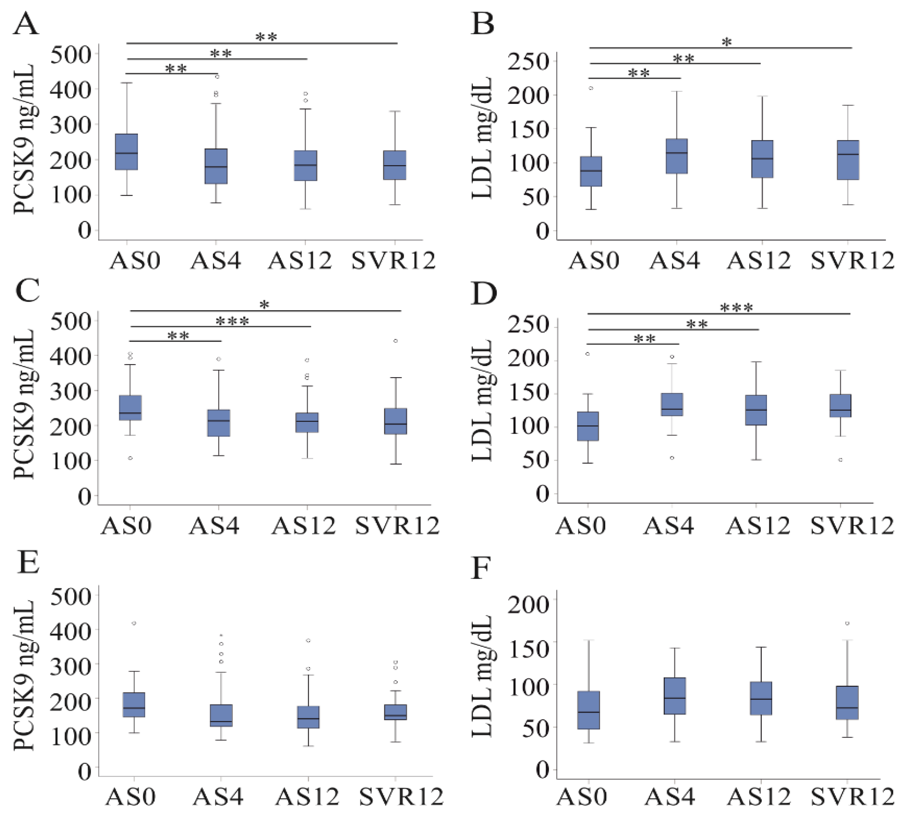
3.4. DAA Therapy Reduces Serum PCSK9 Levels in Non-Cirrhosis Patients

PCSK9 was reduced from 218 (99–418) ng/mL to 179 (78–390) ng/mL already at 4 weeks after the start of DAA therapy in the whole cohort (Figure 4A). Recently, we have shown that LDL levels were induced already at 4 weeks after the start of DAA therapy in this cohort [5], and this is illustrated in a diagram (Figure 4B). Because serum PCSK9 was low in liver cirrhotic patients before therapy start (Figure 2F), DAA-treatment-related effects were separately calculated in HCV patients with and without cirrhosis. Serum PCSK9 was 235 (106–406) ng/mL in non-cirrhotic patients before therapy and 213 (114–390) ng/mL at 4 weeks after therapy start (Figure 4C). In the cirrhotic patients, PCSK9 was 171 (99–418) ng/mL before therapy and 132 (78–383) ng/mL at 4 weeks after therapy start (Figure 4E). The decline of PCSK9 during DAA therapy was significant in non-cirrhotic patients (Figure 4C). LDL was induced in non-cirrhotic patients at all of the time points (Figure 4D). In HCV patients with liver cirrhosis, DAA therapy was not associated with higher LDL levels (Figure 4F).
At all of the time points, serum PCSK9 levels were higher in non-cirrhotic than in cirrhotic patients (p < 0.001 for 4 and 12 weeks after therapy start and for 12 weeks posttreatment).
At 12 weeks posttreatment, the associations of serum PCSK9 with the MELD score, bilirubin, INR, leukocytes and CRP in the patients with liver cirrhosis persisted. At this time point, a positive correlation of PCSK9 and LDL was observed (Table 4).
A poor correlation of serum PCSK9 with albumin was detected in non-cirrhotic patients at 12 weeks posttreatment (Table 4).

4. Discussion

Current analysis showed a weak association of serum PCSK9 with LDL in patients with chronic HCV infection. PCSK9 was low in patients with liver cirrhosis and correlated with measures of liver injury. In non-cirrhotic HCV patients, serum PCSK9 was positively associated with viral load. Serum PCSK9 declined during DAA therapy in non-cirrhotic but not cirrhotic HCV patients. In cirrhotic patients, the impact of liver disease seems to override the effect of viral eradication on serum PCSK9.
Few researchers have compared PCSK9 levels in HCV-infected patients and non-infected controls. Bridge et al. showed that HCV genotype 3 patients had lower and HCV genotype 1 patients had higher plasma PCSK9 levels compared to uninfected controls [24]. It must be noted that plasma and serum PCSK9 levels do not correlate, and it is debatable whether studies using different biofluids can be compared [34]. In HIV-infected patients, coinfection with HCV increased plasma PCSK9 levels [35]. Fasolato et al. observed higher plasma PCSK9 in HCV-infected patients than in non-infected controls [22]. Our study identified elevated PCSK9 levels in serum of patients with chronic HCV compared to non-infected patients without severe liver diseases. This also applies to patients with liver cirrhosis where PCSK9 was higher in HCV patients than patients with non-viral disease etiology. Thus, HCV infection seems to induce serum and plasma PCSK9 and, when comparing different patient cohorts, the severity of liver fibrosis must be considered
PCSK9 declined during prolonged fasting, while levels were not reduced after fasting for 8 to 12 h [36]. Serum PCSK9 levels were moreover induced by dietary saturated fat, fructose and cholesterol [37]. The studies cited above regarding systemic PCSK9 levels in HCV-infected patients used fasting plasma [22,24,35]. Fasting serum was obtained from the patients in the current analysis. A diurnal rhythm, with the lowest PCSK9 levels between 3 and 9 p.m. and a peak at about 4:30 a.m., was also reported [38]. Samples of our cohort were all obtained in the morning, and this may also apply to the studies cited above [22,24,35].
Lipid-lowering drugs were shown to affect circulating PCSK9 levels. Statins increase hepatocyte LDL-receptor and PCSK9 expression, and previous studies described that statins induced systemic PCSK9 concentrations by 6 to 39% [39]. Patients enrolled in the study by Fasolato et al. did not receive statins [22], and statins were temporally discontinued in our patient cohort. The patients analyzed in the study of Kohli et al. and Bridge et al. were not preselected for cardiovascular risk factors, and the use of lipid-lowering drugs was not reported [24,35]. There is some evidence that ezetimibe may also induce PCSK9, whereas fibrates seem to lower its levels [39]. Intake of these drugs is mostly not discontinued during DAA therapy, and the effects of these drugs on circulating PCSK9 may be similar before and after viral eradication. Therefore, it is unlikely that DAA therapy-induced changes of PCSK9 are associated with these medications.
About 18% of the US population was on statin therapy in 2014 [40], and the prevalence of statin use seems to be similar in HCV-infected patients [41]. Thus, higher levels of serum PCSK9 in HCV patients are most likely not a consequence of statin therapy.
HCV genotype appeared to modulate plasma PCSK9 levels in a previous cohort [24] but was not associated with serum PCSK9 in the current analysis. Prevalence of liver cirrhosis and MELD scores did not vary between the genotypes in the present study. The MELD score was not reported for the patients enrolled by Bridge et al. [24] and may be a confounding variable. A second study reported comparable PCSK9 levels in patients with genotype 1a, 1b and 2. Genotype 3 patients had lower PCSK9 levels compared to genotype 2-infected patients. The MELD score was similar between these subgroups [22]. Genotype 2 was rare in the current study cohort, and statistical analysis was, therefore, not possible. However, similar levels of PCSK9 in genotype 1a and 1b-infected patients [22] were following the present observations.
Serum PCSK9 positively correlated with albumin and negatively with the MELD score in our HCV patient cohort, and this is in agreement with a previous analysis [22]. These associations existed in the HCV-infected patients with liver cirrhosis but not in the non-cirrhotic group. Associations of PCSK9 and the MELD score were still significant after correcting for confounding variables. Correlations of PCSK9 and the MELD score could not be observed in non-HCV-infected patients with severe liver diseases [22]. Plasma PCSK9 was not changed with increasing MELD score in liver cirrhotic patients with mostly alcoholic disease etiology [10]. Schlegel et al. nevertheless described a negative correlation of serum PCSK9 with the MELD score in a cohort of patients with severe liver disease and mixed disease etiology. Notably, only 2.7% of these patients were infected with HCV [42]. This suggests that associations of PCSK9 with residual liver function are affected by liver disease severity and disease etiology.
Hepatic PCSK9 protein expression was found increased in the cirrhotic liver, and hepatocellular carcinoma adjacent tissues were used in this analysis. PCSK9 was determined by immunohistochemistry; the fibrosis stage could have been evaluated by histopathology. This study did, however, not discriminate between HCV-infected patients and patients with other disease etiologies [43]. A separate study also described higher PCSK9 mRNA and protein in the fibrotic human liver obtained from patients with hepatitis B infection and patients with non-viral disease etiology [44].
Associations of hepatic PCSK9 protein with lobular inflammation and hepatocyte ballooning were, however, not observed in patients with non-alcoholic fatty liver disease (NAFLD) [45]. Serum PCSK9 was nevertheless positively correlated with steatosis grade, necroinflammation, hepatocyte ballooning, and fibrosis stage in the second cohort of patients with NAFLD. Of note, hepatic PCSK9 expression was also associated with liver steatosis. Correlation of systemic PCSK9 and hepatic expression of lipogenic genes suggested that PCSK9 is induced in parallel with de novo lipogenesis [46]. Correlations of serum PCSK9, LDL and triglycerides did not exist in that study group. Of note, there was a highly significant inverse association of serum PCSK9 with BMI in this cohort [46].
Moreover, hepatic PCSK9 protein was not induced in liver cirrhotic patients of a further investigation, and this applied to patients with HCV or HBV infection and patients with non-viral disease etiology [10].
A sample size of studies analyzing hepatic PCSK9 protein expression was rather small. Thus, further research in larger populations must resolve the complex interplay between PCSK9 levels in serum, hepatic synthesis, liver disease severity and disease etiology.
Here, confounding variables like obesity, liver steatosis and obesity must be considered. The role of PCSK9 in glucose homeostasis and diabetes is still controversial [47]. At least in our cohort of HCV patients, serum PCSK9 was not induced in patients with diabetes. PCSK9 did not correlate with BMI in the HCV patients. Separate studies reported negative and positive associations between PCSK9 and BMI [46,48]. Serum PCSK9 was not induced in HCV patients with liver steatosis but was higher in patients with metabolic fatty liver [46]. These contradictory results show that the association of PCSK9 with metabolic traits is far from being resolved.
Inflammation in HCV infection has a critical role in liver disease progression [49]. Correlations of PCSK9 with CRP and leukocyte count were described before [47]. PCSK9 expression was enhanced in inflammation, and subsequent degradation of the hepatic LDL-receptor hindered the clearance of bacterial lipids, such as lipopolysaccharide [47]. In our study, PCSK9 was positively correlated with leukocyte count in cirrhotic patients. Interestingly, a negative association of CRP and PCSK9 existed in HCV-infected patients with cirrhosis before therapy and at 12 weeks posttreatment. CRP is produced by the liver as an acute-phase protein, and impaired IL-6 signaling, lower hepatocyte synthesis and increased inflammation may regulate CRP levels in HCV infection [50]. Inhibitory monoclonal PCSK9 antibodies did not reduce highly sensitive CRP levels in patients with an increased risk for cardiovascular diseases [51]. Infusion of endotoxin-induced CRP, but not PCSK9 in healthy volunteers [52]. These findings demonstrate that PCSK9 levels are not directly related to CRP [51].
Serum PCSK9 positively correlated with viral load in the subgroup of non-cirrhotic patients confirming a previous study where a positive correlation of HCV viral titer and PCSK9 levels was described [22]. This suggests that HCV may induce serum PCSK9 levels, and HCV infection indeed enhanced hepatic PCSK9 protein [10]. Accordingly, highly efficient eradication of HCV by DAA therapy was linked with a reduction of serum PCSK9 levels already four weeks after the start of treatment. This is in line with a further analysis, where total PCSK9 levels declined four weeks after the start of DAA treatment [27]. In contradiction to these findings, a separate study reported on a rise of PCSK9 in HCV patients with a sustained virologic response [53]. Patients were treated with interferon-based therapies, which were combined with DAAs for a not further specified number of patients [53]. Thus, the effect of HCV eradication on systemic PCSK9 levels is still an unresolved issue.
A main function of PCSK9 is to regulate LDL, and high PCSK9 is associated with high LDL [54]. Indeed, positive associations of PCSK9, cholesterol, LDL and triglycerides were identified in large cohorts [37]. Triglycerides were not documented in our patient cohort, and this is a limitation of our study. Serum PCSK9 did not correlate with LDL levels in cirrhotic and non-cirrhotic HCV-infected patients. Similarly, there were no associations of plasma PCSK9 and LDL in genotype 3- and 1-infected HCV patients [24]. Levels of active PCSK9 did not correlate with LDL in HCV patients [27]. Liver function is the main regulator of serum cholesterol levels, and impaired hepatic lipoprotein release seems to be causal for low LDL levels in patients with severe liver diseases [23]. There was, however, a modest positive correlation between PCSK9 and LDL in cirrhotic HCV patients at 12 weeks posttreatment. Such an association was not observed in the non-cirrhotic patients. Whether the functional role of PCSK9 in LDL metabolism was restored in severely ill patients after HCV elimination needs further research.
High LDL is a risk factor for cardiovascular diseases, and statins are commonly used drugs to treat hypercholesterinemia [55,56]. Statins are well tolerated in patients with compensated liver cirrhosis. In statin-intolerant patients, PCSK9 inhibitors may be used to lower LDL [57]. There are still concerns about the adverse effects of statins in patients with decompensated liver cirrhosis [55,56], and inhibition of PCSK9 may be an alternative approach. Drug–drug interactions of statins and DAAs have been described [56] and PCSK9 inhibitors may be better suited during DAA therapy [57]. Considering the anti-inflammatory and antifibrotic effects of PCSK9 inhibition, blockage of PCSK9 may have multiple beneficial effects in patients with liver cirrhosis [44,57]. Statins also decrease liver cirrhosis incidence [58], and further studies must evaluate whether inhibition of PCSK9 is of advantage. There is some evidence for the role of PCSK9 in HCV infection [18,20], and PCSK9 inhibitors may have antiviral effects. Whether serum PCSK9 levels are associated with chronic HCV infection risk must be evaluated in the future. The rapid decline of PCSK9 after viral elimination suggests a direct effect of the virus on PCSK9 serum levels. The underlying pathways and the role of liver cirrhosis on the crosstalk between HCV and PCSK9 levels must be evaluated in detail.
The study’s limitation was the small sample size, and the association of viral genotype and serum PCSK9 needs further studies. Regarding lipids, this study focused on LDL levels, and correlations with further lipoproteins, fatty acids or triglycerides were not evaluated. Moreover, the ELISA used did not discriminate between active and inactive PCSK9. Study strength was the comparison of HCV-infected and non-infected cohorts. Moreover, serum PCSK9 was measured at different time points during DAA therapy.

5. Conclusions

Positive associations of serum PCSK9 with viral load and decline after viral eradication in non-cirrhotic HCV patients indicate a role of HCV infection to regulate serum PCSK9. In HCV patients with liver cirrhosis, PCSK9 was low and was associated with markers of liver injury. PCSK9 levels did, accordingly, not decline in patients with liver cirrhosis after DAA therapy, which cannot improve liver function within a short time of observation. PCSK9 and LDL were changed in the opposite direction after DAA therapy. Thus, PCSK9 did not contribute to the recovery of LDL levels after the elimination of HCV. Liver function and HCV infection seem to affect serum PCSK9 and LDL levels. Thus, LDL and PCSK9 normalize after viral elimination in patients with “good” liver function. In cirrhotic patients, the impact of impaired liver function may override the effects of viral eradication.

Supplementary Materials

Author Contributions

Conceptualization, K.W., G.P. and C.B.; resources, K.W., G.P., D.S., R.W. and J.G.; investigation, J.G.; statistical analysis, C.B.; writing—original draft preparation, C.B.; writing—review and editing, K.W., G.P., J.G., D.S., R.W., C.B. and M.M., supervision, K.W. and G.P. All authors have read and agreed to the published version of the manuscript.

Funding

There was no external support for this study.

Institutional Review Board Statement

The study was approved by the local ethical committee of the University Hospital of Regensburg (protocol code: 14-101-0049; date of approval: 22 May 2014) and was performed according to the updated guidelines of good clinical practice.

Informed Consent Statement

Informed consent was obtained from all patients involved in the study.

Data Availability Statement

The data presented in this study are available on request from the corresponding author.

Acknowledgments

The expert technical assistance of Elena Underberg is greatly appreciated.

Conflicts of Interest

The authors declare no conflict of interest.

References

  1. Casiraghi, M.A.; De Paschale, M.; Romano, L.; Biffi, R.; Assi, A.; Binelli, G.; Zanetti, A.R. Long-term outcome (35 years) of hepatitis C after acquisition of infection through mini transfusions of blood given at birth. Hepatology 2004, 39, 90–96. [Google Scholar] [CrossRef] [PubMed]
  2. Pawlotsky, J.M.; Negro, F.; Aghemo, A.; Berenguer, M.; Dalgard, O.; Dusheiko, G.; Marra, F.; Puoti, M.; Wedemeyer, H. EASL recommendations on treatment of hepatitis C: Final update of the series. J. Hepatol. 2020, 73, 1170–1218. [Google Scholar] [CrossRef] [PubMed]
  3. Li, J.; Wu, D.B.; Jiang, W.; Chen, X.B.; Xiao, G.B.; Wang, Y.H.; Wang, M.L.; Tao, Y.C.; Chen, E.Q. Efficacy and safety of sofosbuvir-based pangenotypic direct-acting antiviral agents for chronic hepatitis C patients without genotype determination: Real-world experience of a retrospective study. Medicine 2020, 99, e22726. [Google Scholar] [CrossRef]
  4. Nakajima, T.; Karino, Y.; Hige, S.; Suii, H.; Tatsumi, R.; Yamaguchi, M.; Arakawa, T.; Kuwata, Y.; Hasegawa, T.; Toyota, J. Factors affecting the recovery of hepatic reserve after sustained virologic response by direct-acting antiviral agents in chronic hepatitis C virus-infected patients. J. Gastroenterol. Hepatol. 2020, 36, 367–375. [Google Scholar] [CrossRef] [PubMed]
  5. Peschel, G.; Grimm, J.; Gulow, K.; Muller, M.; Buechler, C.; Weigand, K. Chemerin Is a Valuable Biomarker in Patients with HCV Infection and Correlates with Liver Injury. Diagnostics 2020, 10, 974. [Google Scholar] [CrossRef] [PubMed]
  6. Hasan, Y.; Brown, K. Viral eradication restores normal iron status in chronic hepatitis C patients with abnormal iron studies. Ann. Hepatol. 2020, 19, 422–426. [Google Scholar] [CrossRef]
  7. Verna, E.C.; Morelli, G.; Terrault, N.A.; Lok, A.S.; Lim, J.K.; Di Bisceglie, A.M.; Zeuzem, S.; Landis, C.S.; Kwo, P.; Hassan, M.; et al. DAA therapy and long-term hepatic function in advanced/decompensated cirrhosis: Real-world experience from HCV-TARGET cohort. J. Hepatol. 2020, 73, 540–548. [Google Scholar] [CrossRef]
  8. Enomoto, M.; Kawada, N. The Moral of Hepatic Fibrosis: Don’t Always Believe Noninvasive Fibrosis Measurements. Dig. Dis. Sci. 2020, 65, 1293–1295. [Google Scholar] [CrossRef] [Green Version]
  9. Syed, G.H.; Tang, H.; Khan, M.; Hassanein, T.; Liu, J.; Siddiqui, A. Hepatitis C virus stimulates low-density lipoprotein receptor expression to facilitate viral propagation. J. Virol. 2014, 88, 2519–2529. [Google Scholar] [CrossRef] [Green Version]
  10. Feder, S.; Wiest, R.; Weiss, T.S.; Aslanidis, C.; Schacherer, D.; Krautbauer, S.; Liebisch, G.; Buechler, C. Proprotein convertase subtilisin/kexin type 9 (PCSK9) levels are not associated with severity of liver disease and are inversely related to cholesterol in a cohort of thirty eight patients with liver cirrhosis. Lipids Health Dis. 2021, 20, 1–14. [Google Scholar] [CrossRef]
  11. Asselah, T.; Rubbia-Brandt, L.; Marcellin, P.; Negro, F. Steatosis in chronic hepatitis C: Why does it really matter? Gut 2006, 55, 123–130. [Google Scholar] [CrossRef] [PubMed] [Green Version]
  12. Lonardo, A.; Adinolfi, L.E.; Loria, P.; Carulli, N.; Ruggiero, G.; Day, C.P. Steatosis and hepatitis C virus: Mechanisms and significance for hepatic and extrahepatic disease. Gastroenterology 2004, 126, 586–597. [Google Scholar] [CrossRef]
  13. Hashimoto, S.; Yatsuhashi, H.; Abiru, S.; Yamasaki, K.; Komori, A.; Nagaoka, S.; Saeki, A.; Uchida, S.; Bekki, S.; Kugiyama, Y.; et al. Rapid Increase in Serum Low-Density Lipoprotein Cholesterol Concentration during Hepatitis C Interferon-Free Treatment. PLoS ONE 2016, 11, e0163644. [Google Scholar] [CrossRef]
  14. Zaid, A.; Roubtsova, A.; Essalmani, R.; Marcinkiewicz, J.; Chamberland, A.; Hamelin, J.; Tremblay, M.; Jacques, H.; Jin, W.; Davignon, J.; et al. Proprotein convertase subtilisin/kexin type 9 (PCSK9): Hepatocyte-specific low-density lipoprotein receptor degradation and critical role in mouse liver regeneration. Hepatology 2008, 48, 646–654. [Google Scholar] [CrossRef] [PubMed]
  15. Katzmann, J.L.; Gouni-Berthold, I.; Laufs, U. PCSK9 Inhibition: Insights From Clinical Trials and Future Prospects. Front. Physiol. 2020, 11, 595819. [Google Scholar] [CrossRef] [PubMed]
  16. Santulli, G.; Jankauskas, S.S.; Gambardella, J. Inclisiran: A new milestone on the PCSK9 road to tackle cardiovascular risk. Eur. Heart J. Cardiovasc. Pharm. 2021. [Google Scholar] [CrossRef] [PubMed]
  17. Macchi, C.; Sirtori, C.R.; Corsini, A.; Santos, R.D.; Watts, G.F.; Ruscica, M. A new dawn for managing dyslipidemias: The era of rna-based therapies. Pharmacol. Res. 2019, 150, 104413. [Google Scholar] [CrossRef]
  18. Labonte, P.; Begley, S.; Guevin, C.; Asselin, M.C.; Nassoury, N.; Mayer, G.; Prat, A.; Seidah, N.G. PCSK9 impedes hepatitis C virus infection in vitro and modulates liver CD81 expression. Hepatology 2009, 50, 17–24. [Google Scholar] [CrossRef] [PubMed]
  19. Pirro, M.; Bianconi, V.; Francisci, D.; Schiaroli, E.; Bagaglia, F.; Sahebkar, A.; Baldelli, F. Hepatitis C virus and proprotein convertase subtilisin/kexin type 9: A detrimental interaction to increase viral infectivity and disrupt lipid metabolism. J. Cell. Mol. Med. 2017, 21, 3150–3161. [Google Scholar] [CrossRef] [Green Version]
  20. Li, Z.; Liu, Q. Proprotein convertase subtilisin/kexin type 9 inhibits hepatitis C virus replication through interacting with NS5A. J. Gen. Virol. 2018, 99, 44–61. [Google Scholar] [CrossRef]
  21. Ramanathan, A.; Gusarova, V.; Stahl, N.; Gurnett-Bander, A.; Kyratsous, C.A. Alirocumab, a Therapeutic Human Antibody to PCSK9, Does Not Affect CD81 Levels or Hepatitis C Virus Entry and Replication into Hepatocytes. PLoS ONE 2016, 11, e0154498. [Google Scholar] [CrossRef] [Green Version]
  22. Fasolato, S.; Pigozzo, S.; Pontisso, P.; Angeli, P.; Ruscica, M.; Savarino, E.; De Martin, S.; Lupo, M.G.; Ferri, N. PCSK9 Levels Are Raised in Chronic HCV Patients with Hepatocellular Carcinoma. J. Clin. Med. 2020, 9, 3134. [Google Scholar] [CrossRef] [PubMed]
  23. Buechler, C.; Haberl, E.M.; Rein-Fischboeck, L.; Aslanidis, C. Adipokines in Liver Cirrhosis. Int. J. Mol. Sci. 2017, 18, 1392. [Google Scholar] [CrossRef] [PubMed] [Green Version]
  24. Bridge, S.H.; Sheridan, D.A.; Felmlee, D.J.; Crossey, M.M.; Fenwick, F.I.; Lanyon, C.V.; Dubuc, G.; Seidah, N.G.; Davignon, J.; Thomas, H.C.; et al. PCSK9, apolipoprotein E and lipoviral particles in chronic hepatitis C genotype 3: Evidence for genotype-specific regulation of lipoprotein metabolism. J. Hepatol. 2015, 62, 763–770. [Google Scholar] [CrossRef] [Green Version]
  25. Endo, D.; Satoh, K.; Shimada, N.; Hokari, A.; Aizawa, Y. Impact of interferon-free antivirus therapy on lipid profiles in patients with chronic hepatitis C genotype 1b. World J. Gastroenterol. 2017, 23, 2355–2364. [Google Scholar] [CrossRef] [Green Version]
  26. Han, B.; Eacho, P.I.; Knierman, M.D.; Troutt, J.S.; Konrad, R.J.; Yu, X.; Schroeder, K.M. Isolation and characterization of the circulating truncated form of PCSK9. J. Lipid Res. 2014, 55, 1505–1514. [Google Scholar] [CrossRef] [Green Version]
  27. Ichikawa, T.; Miyaaki, H.; Miuma, S.; Taura, N.; Motoyoshi, Y.; Akahoshi, H.; Nakamura, J.; Takahashi, Y.; Honda, T.; Yajima, H.; et al. Changes in serum LDL, PCSK9 and microRNA-122 in patients with chronic HCV infection receiving Daclatasvir/Asunaprevir. Biomed. Rep. 2019, 10, 156–164. [Google Scholar] [CrossRef] [PubMed] [Green Version]
  28. Kiser, J.J.; Burton, J.R.; Anderson, P.L.; Everson, G.T. Review and management of drug interactions with boceprevir and telaprevir. Hepatology 2012, 55, 1620–1628. [Google Scholar] [CrossRef] [Green Version]
  29. Yen, Y.H.; Kuo, F.Y.; Chen, C.H.; Hu, T.H.; Lu, S.N.; Wang, J.H.; Hung, C.H. Ultrasound is highly specific in diagnosing compensated cirrhosis in chronic hepatitis C patients in real world clinical practice. Medicine 2019, 98, e16270. [Google Scholar] [CrossRef]
  30. Butt, A.A.; Xiaoqiang, W.; Budoff, M.; Leaf, D.; Kuller, L.H.; Justice, A.C. Hepatitis C virus infection and the risk of coronary disease. Clin. Infect. Dis. 2009, 49, 225–232. [Google Scholar] [CrossRef] [Green Version]
  31. Angulo, P.; Hui, J.M.; Marchesini, G.; Bugianesi, E.; George, J.; Farrell, G.C.; Enders, F.; Saksena, S.; Burt, A.D.; Bida, J.P.; et al. The NAFLD fibrosis score: A noninvasive system that identifies liver fibrosis in patients with NAFLD. Hepatology 2007, 45, 846–854. [Google Scholar] [CrossRef] [PubMed]
  32. Ho, S.Y.; Liu, P.H.; Hsu, C.Y.; Hsia, C.Y.; Su, C.W.; He, Y.J.; Lee, Y.H.; Huang, Y.H.; Hou, M.C.; Huo, T.I. Current noninvasive liver reserve models do not predict histological fibrosis severity in hepatocellular carcinoma. Sci. Rep. 2018, 8, 15074. [Google Scholar] [CrossRef] [PubMed]
  33. Ragazzo, T.G.; Paranagua-Vezozzo, D.; Lima, F.R.; de Campos Mazo, D.F.; Pessoa, M.G.; Oliveira, C.P.; Alves, V.A.F.; Carrilho, F.J. Accuracy of transient elastography-FibroScan(R), acoustic radiation force impulse (ARFI) imaging, the enhanced liver fibrosis (ELF) test, APRI, and the FIB-4 index compared with liver biopsy in patients with chronic hepatitis C. Clinics 2017, 72, 516–525. [Google Scholar] [CrossRef]
  34. Rajan, M.R.; Sotak, M.; Barrenas, F.; Shen, T.; Borkowski, K.; Ashton, N.J.; Biorserud, C.; Lindahl, T.L.; Ramstrom, S.; Scholl, M.; et al. Comparative analysis of obesity-related cardiometabolic and renal biomarkers in human plasma and serum. Sci. Rep. 2019, 9, 15385. [Google Scholar] [CrossRef] [PubMed] [Green Version]
  35. Kohli, P.; Ganz, P.; Ma, Y.; Scherzer, R.; Hur, S.; Weigel, B.; Grunfeld, C.; Deeks, S.; Wasserman, S.; Scott, R.; et al. HIV and Hepatitis C-Coinfected Patients Have Lower Low-Density Lipoprotein Cholesterol Despite Higher Proprotein Convertase Subtilisin Kexin 9 (PCSK9): An Apparent “PCSK9-Lipid Paradox”. J. Am. Heart Assoc. 2016, 5, e002683. [Google Scholar] [CrossRef] [PubMed] [Green Version]
  36. Browning, J.D.; Horton, J.D. Fasting reduces plasma proprotein convertase, subtilisin/kexin type 9 and cholesterol biosynthesis in humans. J. Lipid Res. 2010, 51, 3359–3363. [Google Scholar] [CrossRef] [PubMed] [Green Version]
  37. Krysa, J.A.; Ooi, T.C.; Proctor, S.D.; Vine, D.F. Nutritional and Lipid Modulation of PCSK9: Effects on Cardiometabolic Risk Factors. J. Nutr. 2017, 147, 473–481. [Google Scholar] [CrossRef] [Green Version]
  38. Persson, L.; Cao, G.; Stahle, L.; Sjoberg, B.G.; Troutt, J.S.; Konrad, R.J.; Galman, C.; Wallen, H.; Eriksson, M.; Hafstrom, I.; et al. Circulating proprotein convertase subtilisin kexin type 9 has a diurnal rhythm synchronous with cholesterol synthesis and is reduced by fasting in humans. Arter. Thromb Vasc. Biol. 2010, 30, 2666–2672. [Google Scholar] [CrossRef] [Green Version]
  39. Nozue, T. Lipid Lowering Therapy and Circulating PCSK9 Concentration. J. Atheroscler. Thromb. 2017, 24, 895–907. [Google Scholar] [CrossRef] [Green Version]
  40. Bucholz, E.M.; Rodday, A.M.; Kolor, K.; Khoury, M.J.; de Ferranti, S.D. Prevalence and Predictors of Cholesterol Screening, Awareness, and Statin Treatment Among US Adults with Familial Hypercholesterolemia or Other Forms of Severe Dyslipidemia (1999–2014). Circulation 2018, 137, 2218–2230. [Google Scholar] [CrossRef]
  41. Rao, G.A.; Pandya, P.K. Statin therapy improves sustained virologic response among diabetic patients with chronic hepatitis C. Gastroenterology 2011, 140, 144–152. [Google Scholar] [CrossRef] [PubMed]
  42. Schlegel, V.; Treuner-Kaueroff, T.; Seehofer, D.; Berg, T.; Becker, S.; Ceglarek, U.; Thiery, J.; Kaiser, T. Low PCSK9 levels are correlated with mortality in patients with end-stage liver disease. PLoS ONE 2017, 12, e0181540. [Google Scholar] [CrossRef] [PubMed] [Green Version]
  43. Bhat, M.; Skill, N.; Marcus, V.; Deschenes, M.; Tan, X.; Bouteaud, J.; Negi, S.; Awan, Z.; Aikin, R.; Kwan, J.; et al. Decreased PCSK9 expression in human hepatocellular carcinoma. BMC Gastroenterol. 2015, 15, 1–10. [Google Scholar] [CrossRef] [Green Version]
  44. Zou, Y.; Li, S.; Xu, B.; Guo, H.; Zhang, S.; Cai, Y. Inhibition of Proprotein Convertase Subtilisin/Kexin Type 9 Ameliorates Liver Fibrosis via Mitigation of Intestinal Endotoxemia. Inflammation 2020, 43, 251–263. [Google Scholar] [CrossRef]
  45. Emma, M.R.; Giannitrapani, L.; Cabibi, D.; Porcasi, R.; Pantuso, G.; Augello, G.; Giglio, R.V.; Re, N.L.; Capitano, A.R.; Montalto, G.; et al. Hepatic and circulating levels of PCSK9 in morbidly obese patients: Relation with severity of liver steatosis. Biochim. Biophys. Acta Mol. Cell Biol. Lipids 2020, 1865, 158792. [Google Scholar] [CrossRef] [PubMed]
  46. Ruscica, M.; Ferri, N.; Macchi, C.; Meroni, M.; Lanti, C.; Ricci, C.; Maggioni, M.; Fracanzani, A.L.; Badiali, S.; Fargion, S.; et al. Liver fat accumulation is associated with circulating PCSK9. Ann. Med. 2016, 48, 384–391. [Google Scholar] [CrossRef] [PubMed]
  47. Cesaro, A.; Bianconi, V.; Gragnano, F.; Moscarella, E.; Fimiani, F.; Monda, E.; Scudiero, O.; Limongelli, G.; Pirro, M.; Calabro, P. Beyond cholesterol metabolism: The pleiotropic effects of proprotein convertase subtilisin/kexin type 9 (PCSK9). Genetics, mutations, expression, and perspective for long-term inhibition. Biofactors 2020, 46, 367–380. [Google Scholar] [CrossRef]
  48. Toth, S.; Fedacko, J.; Pekarova, T.; Hertelyova, Z.; Katz, M.; Mughees, A.; Kuzma, J.; Stefanic, P.; Kopolovets, I.; Pella, D. Elevated Circulating PCSK9 Concentrations Predict Subclinical Atherosclerotic Changes in Low Risk Obese and Non-Obese Patients. Cardiol. Ther. 2017, 6, 281–289. [Google Scholar] [CrossRef]
  49. Giannelli, G.; Antonaci, S. Immunological and molecular aspects of liver fibrosis in chronic hepatitis C virus infection. Histol. Histopathol. 2005, 20, 939–944. [Google Scholar] [CrossRef]
  50. Babiker, A.; Hassan, M.; Muhammed, S.; Taylor, G.; Poonia, B.; Shah, A.; Bagchi, S. Inflammatory and cardiovascular diseases biomarkers in chronic hepatitis C virus infection: A review. Clin. Cardiol. 2020, 43, 222–234. [Google Scholar] [CrossRef]
  51. Ruscica, M.; Tokgozoglu, L.; Corsini, A.; Sirtori, C.R. PCSK9 inhibition and inflammation: A narrative review. Atherosclerosis 2019, 288, 146–155. [Google Scholar] [CrossRef] [PubMed] [Green Version]
  52. Heinzl, M.W.; Resl, M.; Klammer, C.; Egger, M.; Dieplinger, B.; Clodi, M. Proprotein Convertase Subtilisin/Kexin Type 9 (PCSK9) Is Not Induced in Artificial Human Inflammation and Is Not Correlated with Inflammatory Response. Infect. Immun. 2020, 88. [Google Scholar] [CrossRef] [PubMed] [Green Version]
  53. Hyrina, A.; Olmstead, A.D.; Steven, P.; Krajden, M.; Tam, E.; Jean, F. Treatment-Induced Viral Cure of Hepatitis C Virus-Infected Patients Involves a Dynamic Interplay among three Important Molecular Players in Lipid Homeostasis: Circulating microRNA (miR)-24, miR-223, and Proprotein Convertase Subtilisin/Kexin Type 9. EBioMedicine 2017, 23, 68–78. [Google Scholar] [CrossRef] [PubMed] [Green Version]
  54. Mousavi, S.A.; Berge, K.E.; Leren, T.P. The unique role of proprotein convertase subtilisin/kexin 9 in cholesterol homeostasis. J. Intern. Med. 2009, 266, 507–519. [Google Scholar] [CrossRef] [PubMed]
  55. Kaplan, D.E.; Serper, M.A.; Mehta, R.; Fox, R.; John, B.; Aytaman, A.; Baytarian, M.; Hunt, K.; Albrecht, J.; Njei, B.; et al. Effects of Hypercholesterolemia and Statin Exposure on Survival in a Large National Cohort of Patients With Cirrhosis. Gastroenterology 2019, 156, 1693–1706. [Google Scholar] [CrossRef] [PubMed] [Green Version]
  56. Kuo, M.H.; Tseng, C.W.; Lee, C.H.; Tseng, K.C. Drug-drug interactions between direct-acting antivirals and statins in the treatment of chronic hepatitis C. Tzu Chi Med. J. 2020, 32, 331–338. [Google Scholar] [CrossRef] [PubMed]
  57. Shapiro, M.D.; Tavori, H.; Fazio, S. PCSK9: From Basic Science Discoveries to Clinical Trials. Circ. Res. 2018, 122, 1420–1438. [Google Scholar] [CrossRef]
  58. Wang, Y.; Xiong, J.; Niu, M.; Chen, X.; Gao, L.; Wu, Q.; Zheng, K.; Xu, K. Statins and the risk of cirrhosis in hepatitis B or C patients: A systematic review and dose-response meta-analysis of observational studies. Oncotarget 2017, 8, 59666–59676. [Google Scholar] [CrossRef] [Green Version]
Figure 1. Serum proprotein convertase subtilisin/kexin 9 (PCSK9) in patients with chronic hepatitis C virus (HCV) and controls. (A) Serum PCSK9 in patients with chronic HCV and non-infected controls without severe liver diseases; (B) serum PCSK9 in female and male HCV patients; (C) serum PCSK9 in diabetic and non-diabetic HCV patients; (D) serum PCSK9 in patients stratified for HCV genotype (rare group is 6 patients with genotypes other than 1a, 1b, and 3a). Small circles above the boxes mark outliers. ** p < 0.01.
Jcm 10 01621 g001
Figure 2. Serum PCSK9 in patients with chronic HCV stratified for steatosis, fibrosis scores and cirrhosis. (A) PCSK9 in serum of 37 patients with and 45 patients without liver steatosis; serum PCSK9 levels in patients stratified for fibrosis by (B) the acoustic radiation force impulse (ARFI) score; (C) the fibrosis-4 (FIB-4) score; (D) the non-alcoholic fatty liver disease (NAFLD) score and (E) the aminotransferase/platelet ratio index (APRI) score (no fibrosis = no; not reliable values = NR, fibrosis = yes); (F) serum PCSK9 in 37 patients with liver cirrhosis and 45 non-cirrhotic patients assessed by ultrasound. Small circles above or below the boxes mark outliers. * p < 0.05, ** p < 0.01, *** p < 0.001.
Jcm 10 01621 g002
Figure 3. Serum PCSK9 in controls, non-cirrhotic HCV patients and cirrhotic patients with HCV or non-viral disease etiology. (A) Serum PCSK9 of HCV patients with liver cirrhosis and controls not infected with HCV; (B) Serum PCSK9 of HCV patients without liver cirrhosis and the controls not infected with HCV; (C) Serum PCSK9 in patients with HCV and non-viral liver cirrhosis. Small circles above or below the boxes mark outliers. *** p < 0.001.
Jcm 10 01621 g003
Figure 4. Serum PCSK9 and LDL at 0, 4 and 12 weeks after the start of therapy (AS), and 12 weeks after the end of therapy (SVR12). Serum PCSK9 (A) and LDL (B) in the whole cohort; serum PCSK9 (C) and LDL (D) in non-cirrhotic HCV patients; serum PCSK9 (E) and LDL (F) in cirrhotic HCV patients. Small circles above or below the boxes mark outliers. * p < 0.05, ** p < 0.01 and *** p < 0.001.
Jcm 10 01621 g004
Table 1. Laboratory parameters of the HCV-infected patients and the non-infected control cohort, which included patients without severe liver diseases.
HCVNon-HCVp-Value
Number8255ns
Sex (female/male)33/4923/32ns
Age (years)59 (24–80)58 (21–80)ns
BMI (kg/m2)27.1 (18.4–40.4)26.3 (17.8–39.7)ns
MELD score8 (6–20)na
ALT (U/L)86 (22–287)32 (16–288) 54<0.001
AST (U/L)75 (17–1230)24 (12–256) 54<0.001
Albumin (g/L)36 (19–45) 8147 (32–56)<0.001
Bilirubin (mg/dL)0.8 (0.3–4.3)0.5 (0.2–2.8) 53<0.001
INR1.13 (0.91–2.44)nd
Creatinine (mg/dL)
Ferritin (ng/mL)
0.78 (0.14–1.31) 81
141 (7.0–2309) 76
nd
nd
Leukocytes (n/L)5.9 (2.2–72.4)nd
CRP (mg/L)2.9 (2.9–72.4)nd
LDL (mg/dL)88 (31–210) 77112 (44–340)<0.01
HDL (mg/dL)51 (19–103) 77nd
Viral load (U/mL)1.4 × 106na
Genotype 1a/1b/3a/rare24/38/14/6na
FIB-4 (no fibrosis/intermediate values/yes)14/15/53na
APRI (no fibrosis/no reliable values/fibrosis/cirrhosis)13/12/29/28na
ARFI
(F1/F3/F4)
15/25/42na
NAFLD
(no fibrosis/no reliable values/fibrosis/nd)
17/22/35/8na
PCSK9 (ng/mL)218 (99–418)170 (40–360)<0.01
ALT: alanine aminotransferase; ARFI: acoustic radiation force impulse; AST: aspartate aminotransferase; BMI: body mass index; CRP: C-reactive protein; HCV: hepatitis C virus; HDL: high-density lipoprotein; INR: international normalized ratio; LDL: low-density lipoprotein; NAFLD: non-alcoholic fatty liver disease; na: not applicable; nd: not defined; ns: not significant; PCSK9: proprotein convertase subtilisin/kexin 9; In case that the laboratory values were not documented for the whole cohort, the respective numbers are given in superscript.
Table 2. Laboratory parameters of the cirrhotic patients with HCV and non-viral disease etiology. *** p < 0.001.
HCV CirrhosisNon-HCV Cirrhosis
Number3726
Sex (female/male)15/225/21
Age (years)61 (38–80)49 (40–81)
MELD score10 (7–20)8 (6–21) 22
Albumin (g/L)34 (19–45)32 (2–4)
Bilirubin (mg/dL)1.2 (0.4–4.3)1.1 (0.4–3.7)
LDL (mg/dL)
PCSK9 (ng/mL)
68 (31–152)
171 (99–418)
35 (17–81)
108 (41–290) ***
In case that the laboratory values were not documented for the whole cohort, the respective number is given in superscript.
Table 3. Spearman correlation coefficients and p-values for the correlations of PCSK9 with body mass index (BMI), age, model for end-stage liver disease (MELD) score, viral load and routine laboratory parameters in the HCV patients before direct-acting antiviral (DAA) therapy, at 4 and 12 weeks after the start of therapy and 12 weeks posttreatment.
ParameterBaseline
(82 Patients)
4-Weeks
(79 Patients)
12-Weeks
(81 Patients)
12 Weeks Posttreatment
(76 Patients)
BMI, kg/m20.042 (0.720)0.037 (0.756)0.001 (0.995)−0.026 (0.840)
Age−0.248 (0.024)−0.336 (0.002)−0.180 (0.108)−0.153 (0.221)
MELD score−0.623 (<0.001)−0.579 (<0.001)−0.573 (<0.001)−0.570 (<0.001)
ALT, U/L−0.021 (0.853)−0.042 (0.711)−0.082 (0.467)−0.060 (0.627)
AST, U/L−0.260 (0.018)−0.258 (0.022)−0.288 (0.009)−0.339 (<0.004)
Bilirubin, mg/dL−0.506 (<0.001)−0.587 (<0.001)−0.597 (<0.001)−0.568 (<0.001)
Albumin, g/L0.397 (<0.001)0.460 (<0.001)0.527 (<0.001)0.441 (<0.001)
INR−0.595 (<0.001)−0.549 (<0.001)−0.608 (<0.001)−0.574 (<0.001)
Creatinine, mg/dL0.074 (0.512)−0.013 (0.909)0.010 (0.932)0.159 (0.191)
Leukocytes, n/L0.389 (<0.001)0.358 (0.001)0.503 (<0.001)0.383 (0.001)
CRP, mg/L
HDL, mg/dL
LDL, mg/dL
−0.288 (0.009)
0.047 (0.686)
0.502 (<0.001)
0.218 (0.052)
0.042 (0.720)
0.513 (<0.001)
−0.235 (0.036)
−0.048 (0.674)
0.578 (<0.001)
0.018 (0.885)
0.022 (0.862)
0.595 (<0.001)
Viral load0.298 (0.007)nanana
Significant correlations are in bold. Laboratory parameters were documented for at least 95% of the patients; na: not applicable.
Table 4. Spearman correlation coefficients and p-values (in brackets) for the correlations of PCSK9 with age, MELD score, viral load and routine laboratory parameters in the HCV patients with and without liver cirrhosis before direct-acting antiviral (DAA) therapy, and 12 weeks posttreatment.
ParameterBaseline
(No Cirrhosis; 45 Patients)
Baseline
(Cirrhosis; 37 Patients)
12 Weeks Posttreatment
(No Cirrhosis; 45 Patients)
12 Weeks Posttreatment
(Cirrhosis; 37 Patients)
Age−0.010 (0.949)−0.300 (0.071)−0.168 (0.306)−0.021 (0.913)
MELD score−0.061 (0.689)−0.707 (<0.001)−0.195 (0.234)−0.687 (<0.001)
ALT, U/L−0.019 (0.900)−0.332 (0.045)−0.034 (0.836)−0.009 (0.964)
AST, U/L0.063 (0.681)−0.011 (0.951)0.047 (0.776)−0.323 (0.082)
Bilirubin, mg/dL−0.097 (0.525)−0.653 (<0.001)−0.294 (0.069)−0.603 (0.001)
Albumin, g/L−0.117 (0.451)0.442 (0.006)−0.322 (0.049)0.283 (0.130)
INR−0.083 (0.589)−0.567 (<0.001)−0.180 (0.274)−0.567 (<0.001)
Leukocytes, n/L−0.052 (0.735)0.495 (0.002)−0.083 (0.615)0.367 (0.050)
CRP, mg/L
LDL, mg/dL
−0.185 (0.223)
0.207 (0.193)
−0.526 (0.001)
0.316 (0.061)
−0.218 (0.182)
0.270 (0.123)
−0.526 (0.001)
0.459 (0.014)
Viral load0.326 (0.029)−0.003 (0.987)nana
Laboratory parameters were documented for at least 95% of the patients. Significant correlations are in bold (not applicable, na).
Publisher’s Note: MDPI stays neutral with regard to jurisdictional claims in published maps and institutional affiliations.

