
Chitosan Nanoparticle-Based System: A New Insight into the Promising Controlled Release System for Lung Cancer Treatment


1
Department of Cell and Molecular Biology, Faculty of Biotechnology and Biomolecular Sciences, Universiti Putra Malaysia, Serdang 43400, Selangor, Malaysia
2
UPM-MAKNA Cancer Research Laboratory, Institute of Biosciences, Universiti Putra Malaysia, Serdang 43400, Selangor, Malaysia
*
Author to whom correspondence should be addressed.
Molecules 2022, 27(2), 473; https://0-doi-org.brum.beds.ac.uk/10.3390/molecules27020473
Submission received: 25 February 2021
/
Revised: 15 March 2021
/
Accepted: 25 March 2021
/
Published: 12 January 2022
(This article belongs to the Special Issue Recent Advances in Nanomedicine and Nanovaccine Development)
Abstract
:Lung cancer has been recognized as one of the most often diagnosed and perhaps most lethal cancer diseases worldwide. Conventional chemotherapy for lung cancer-related diseases has bumped into various limitations and challenges, including non-targeted drug delivery, short drug retention period, low therapeutic efficacy, and multidrug resistance (MDR). Chitosan (CS), a natural polymer derived from deacetylation of chitin, and comprised of arbitrarily distributed β-(1-4)-linked d-glucosamine (deacetylated unit) and N-acetyl-d-glucosamine (acetylated unit) that exhibits magnificent characteristics, including being mucoadhesive, biodegradable, and biocompatible, has emerged as an essential element for the development of a nano-particulate delivery vehicle. Additionally, the flexibility of CS structure due to the free protonable amino groups in the CS backbone has made it easy for the modification and functionalization of CS to be developed into a nanoparticle system with high adaptability in lung cancer treatment. In this review, the current state of chitosan nanoparticle (CNP) systems, including the advantages, challenges, and opportunities, will be discussed, followed by drug release mechanisms and mathematical kinetic models. Subsequently, various modification routes of CNP for improved and enhanced therapeutic efficacy, as well as other restrictions of conventional drug administration for lung cancer treatment, are covered.
1. Introduction
Over the last decade, the incidence and mortality rate of cancer has remained one of the leading causes of death worldwide, second to cardiovascular diseases [1]. Based on the analysis of the World Health Organization (WHO), every sixth death in the world is due to cancer [2]. Approximately 1.8 million new lung cancer cases are discovered annually, and about 1.6 million deaths were reported among them, with 4%–17% 5-year survival rates liable on stage and regional difference [3,4,5]. This incidence rate is highly related to the uptake and consumption of tobacco according to gender and habits of people in different geographical areas [6]. Lung cancer can be classified into two main categories, small cell lung carcinoma (SCLC) that accounts for about 15% of all lung cancer, and non-small cell lung carcinoma (NSCLC), accounting for the other 85%. SCLC is known as an aggressive lung tumor that is heavily related to cigarette smoking, with patients often diagnosed with metastatic disease [7]. NSCLC is further sub-divided into adenocarcinoma (most common cases), squamous cell carcinoma, and large cell carcinoma [8]. Adenocarcinoma is most common in NSCLC, which arises from small airway epithelial cells, type II alveolar cells, which secrete mucus and other substances, while squamous-cell carcinoma arises from squamous cells in the airway epithelial cells in the bronchial tubes in the center of the lungs, and large cell carcinoma often arises in the central part of the lung [9]. Researchers have formulated several means of therapy such as surgery, radiation, and chemotherapy to treat cancer patients, but chemotherapy remains the most significant treatment among these approaches, with synthetic drugs such as anthracyclines being the most potent chemotherapeutic drugs used [10]. The four most prominent anthracyclines are epirubicin, doxorubicin, idarubicin, and daunorubicin [11]. Yet, the severe side effects following the chemotherapy due to non-specific destruction have become the main constrain for the treatment, including cardiotoxicity, nausea, and alopecia [12,13].
Arising from these issues, research focus has shifted to the utilization of nanotechnology to increase treatment efficacy and increase treatment safety. Polymeric nanoparticles are a large family which can generally be divided into natural and synthetic forms. The term polymer can be defined as macromolecules that are made up of bulky repeating units arranged in a chain-like structure, presenting an assortment of alignments, structures, and characteristics [14,15]. Natural polymers are comprised of two major types, polysaccharides- and protein-based polymers, whereas synthetic polymers include polyethers, polyesters, poloxamers, and recombinant protein-based polymer [14,16]. Although synthetic polymers have greater flexibility in terms of structure rationalization, natural polymers such as hyaluronic acid, chitosan, and alginate are still more desirable due to their biodegradability and biocompatibility, they are inexpensive and able to be modified [17,18,19]. Consequently, the utilization of chitosan nanoparticle (CNP) systems has been widely recognized by researchers due to their various advantageous characteristics, including being biocompatible, having low toxicity, are easy to prepare, and have tunable physical properties [20,21]. CS is one of the most prominent natural polyaminosaccharides acquired through the N-deacetylation of chitin, the second most abundant natural occurring polysaccharide after cellulose [22,23,24]. The structure of CS is composed of β-(1-4)-linked d-glucosamine and N-acetyl-d-glucosamine randomly disseminated within the polymer, enabling its cationic nature [25,26,27,28]. Besides, CS also has several features such as good biocompatibility and biodegradability, non-toxicity, and low allergenicity, which have fascinated researchers, and allow them to be utilized for numerous applications. Moreover, it was reported that CS possessed several biological properties such as anti-oxidant, anti-microbial, and anti-tumor activities [29,30]. However, there are numerous challenges of using these nanoparticles in anticancer treatments, such as their biocompatibility and toxicity in biological systems, the high attrition of anti-cancer agents in clinical trials, the slow progress of nanomedicines therapeutics to market, technical and cost involved in up-scaling and manufacturing, and poor understanding about the heterogeneity of disease in the patient population [31,32].
The CNP-based nanoparticles in drug delivery have been utilized in various diseases treatment, including cancer-related diseases, gastrointestinal diseases, pulmonary diseases, ocular infections, and drug delivery across the blood-brain barrier [33,34,35,36,37]. CNP possessed several features that enabled its utilization as a vital drug delivery vector, such as enhance delivery and therapeutic efficacy of drugs [38,39], improved intracellular accumulation [40,41], and controlled release properties [42]. Similar to other nanoparticulate delivery systems, CNP has a protective layer after the encapsulation of therapeutics into the core of nano-particles. The conventional methods of drug consumption such as oral delivery, sublingual delivery, and rectal delivery are always preferred by patients due to the ease of consumption widespread acceptance by patients [43]. However, the therapeutic efficacy of these medications is often less desired as the drugs that are consumed, especially orally, need to go through many different paths such as the digestive tract, gastrointestinal tract, and liver to reach the target site, which may lead to the cleavage or degradation by the enzymes or extreme pH environment before they reach the site of absorption and bloodstream [44,45]. Besides that, many drugs may become insoluble at low pH levels in the digestive tract, which may significantly reduce the bioavailability of the drugs to be absorbed into the bloodstream [46]. In order to overcome the obstacles faced by conventional methods, nano-mediated drug delivery systems such as CNPs have received more attention as a delivery system for drugs. The mucoadhesive properties of CNPs also contribute to the absorption and enhanced intracellular accumulation of drugs delivered by CNPs. As mentioned by Tiyaboonchai, both in vitro and in vivo experiments found that CNPs can enhance absorption through the combination of mucoadhesion and transient opening of tight junctions of the mucosal membrane [47]. The interaction between cationic CS and anionic mucin has been attributed to the mucoadhesive properties of CS, which elongates the contact time between the encapsulated drugs and the absorptive surface, and subsequently prolongs the half-life of drug clearance, which in turn enhanced the absorption [48]. Besides that, reports in the literature also show that positively charged CNPs exhibit electrostatic interactions with negatively charged cell membranes, thus revealing a greater uptake through the endo-cytosis pathway [49,50].
The study of Hassan et al. also demonstrated that CNPs promote intracellular accumulation by observing fluorescently-labeled glutamic acids (GA) with and without encapsulation by CNP under a fluorescence microscope. The results showed that the fluorescence signal was detected only in cells that were treated with with fluorescein isothiocyanate (FITC) labeled-GA (FITC-GA) encapsulated CS nanoparticles and was found mainly accumulated in the cell cytoplasm, while no fluorescence signal was detected in cells treated with free FITC-GA [51]. Additionally, CNP as a prominent drug delivery system has been reported to successfully encapsulate various therapeutics from chemotherapeutic drugs, such as doxorubicin, paclitaxel, and carboplatin, to natural anti-cancer compounds such as curcumin and hydrocaffeic acid [52,53,54,55,56,57]. As reported by Zare et al., doxorubicin encapsulated CNP was able to achieve nearly 90% of intestinal permeation post oral administration to the rat model [53]. Besides, Xu et al. has conducted an experiment that described the CNP encapsulation of paclitaxel to study the anti-cancer efficacy against DU-145 prostate cancer cell lines indicated increased growth inhibition effects with an increased concentration of paclitaxel [54]. Khan et al. also described the curcumin encapsulated CNP had enhanced water solubility, bioavailability, and cytotoxic efficacy against various cancer cell lines as compared with free curcumin [58]. On the other hand, Nashaat et al. utilized CNPs for the oral delivery of albumin. An in vivo experiment found that administration of albumin-loaded CNPs had greater enzymatic stability, sustained release, and higher serum concentration as compared with the free albumin [59]. The examples mentioned above have suggested CNP as a promising nano-carrier of anti-cancer therapeutics. Therefore, this review highlights multiple applications of CNP-based systems and their controlled release properties for delivering various therapeutic agents to attain high loading, enhanced therapeutic efficacy, pro-longed retention time, and overcome multidrug resistance (MDR) as effective lung cancer treatment modalities.
2. Current State of Chitosan Nanoparticles in Various Fields of Application
2.1. Advantages
The utilization of nanoparticles for the encapsulation of cargos such as various therapeutic drugs or compounds and genetic materials has been reported by many researchers over the years. As shown in Figure 1, for example, the brief mechanism of a therapeutic-encapsulated CNP system for lung cancer treatment has been illustrated. CS is one of the most prominently used natural polyaminosaccharides due to its characteristics, including good biocompatibility and biodegradability, ease of synthesis, non-toxicity, non-immunogenic, and applicable to a wide range of therapeutics [20,60,61,62]. Besides, as compared with other biodegradable polymers having a pharmacopeial monograph, CS is known as the only polymer that exhibits a cationic character, which resulted in its utilization as a drug delivery system [63]. Additionally, CS was known to possess inhibitory effects on proliferation of tumor cells, tumor-related angiogenesis, and metastasis, and thus demonstrating good anticancer activity [64]. Due to these advantages, it has fascinated the researchers to CNP in numerous applications. Previous toxicology evaluation study of CNP was conducted by Wang et al. on the embryonic development of zebrafish, and it was found that the zebrafish treated by CNPs with an average size of 84.86 nm had no significant mortality (<10%) at 120 h post-fertilization (hpf), even at the high concentration of 200 mg/L [65]. Additionally, this result was supported by another organ-specific toxicology study on zebrafish embryos by Abou-Saleh et al. They reported a 100% survival rate of embryos with no morphological and physiological abnormalities were observed after exposure to CNPs at 200 mg/L at 96 hpf [66]. Apart from that, CNPs have been reported to enhance the therapeutic efficacy of therapeutics, especially in tumor therapy, through passive targeting or enhanced permeation and retention (EPR) effects [67,68,69]. It has been suggested by the previous study of Ai et al. where copper (CuSO4) loaded CNPs showed greater therapeutic efficacy than free CuSO4 groups against osteosarcoma assessed through in vitro cytotoxicity assay, reactive oxygen species (ROS) analysis, and caspase 3 and 7 apoptotic activities analysis due to better internalization of CNP system [70]. On the other hand, On et al. also developed a non-toxic indocyanine green (ICG) and cyanine 5.5 (Cy5.5)-coupled tumor-targeting CNP-based system, which displayed outstanding tumor accumulation and prolonged biodistribution profiles in both rabbit squamous cancer cell (VX2) tumor-bearing mouse and rabbit models probably due to the EPR effects of CNP [71].
Moreover, it was reported that CS and CS derivatives possessed several biological properties such as anti-oxidant [72,73,74], anti-microbial [75,76,77], anti-inflammatory [78,79,80], and anti-tumor activities [81,82,83]. The anti-oxidation properties of CS were suggested to be closely related to its degree of deacetylation and molecular weight, where a lower molecular weight CS was more advantageous than higher molecular weight CS in the removal of free radicals while highly deacetylated CS had superior antioxidant activity compared to less deacetylated CS [84,85,86]. It was supported by previous studies that the free radical scavenging activity of CS may be attributed to the structure of CS with amino and hydroxyl groups attached to the C-2, C-3, and C-6 positions of the pyranose ring of CS, and higher radical elimination activity due to the presence of the high number of amino groups in highly deacetylated CS [87,88,89]. Additionally, the anti-microbial properties of CS have largely contributed to industry applications, especially in wound dressing, food preservation, and pollutants control [77,90]. In food preservation, CS has been widely studied to be used as edible food coating films to acts as an oxygen and low water vapor barrier for fruits and vegetables, extending their shelf life, and at the same time, reducing microorganism growth [91,92]. However, in the context of pollutant removal, Sun et al. proposed that CS had enhanced the antibacterial filter materials for ventilation applications against Escherichia coli showing its decent antimicrobial properties [93].
It has been further shown by a previous study of Al-Sherbini et al. that the development of CS/silver bionanocomposites has aided in photodegradation of organic pollutants, adsorption of heavy metals pollutants, and also showed antibacterial activities on both gram-negative (E. coli) and gram-positive bacteria ( gram positive. bacillus) [94].
2.2. Challenges
However, apart from the advantages of CS and its derivatives in various fields as mentioned above, there are numerous challenges remaining to be resolved to improve its use in the future. One of the main concerns is about the safety of CS and its derivatives, especially in biomedical applications. Generally, CS has been categorized as relatively safe due to its biodegradability and biocompatibility properties. It has been reported that elimination of CS was low molecular weight CS can be excreted through the kidney, while the high molecular form can be renally excreted after degraded into fragments [95]. Nevertheless, the utilization of the native form of CS is restricted, especially in wound management applications, mainly due to its water insolubility, high viscosity, and tendency to coagulate with proteins at high pH values [96]. Besides, Hu et al. has reported concentration-dependent toxicity of CNPs in zebrafish embryos [97]. It has also been suggested by the early study of Huang et al. that CNPs exhibited a cytotoxicity effect at concentrations higher than 0.741 mg/mL [98]. However, it was described by Sonin et al. that an extended 14 days of CNPs treatment in rats did not reveal any significant toxicity effects as assessed by hematological and biochemical parameters of the blood [99]. It has therefore inferred that the toxicity of CS might be due to the size of the CNPs and also the usage of different types of crosslinkers [100,101,102]. Glutaraldehyde, for example, is one of the highly effective cross-linking agents but is toxic to biological systems [103,104]. Besides, Parhi has also revealed that crosslinkers such as glutaraldehyde and glyoxal can potentially initiate toxicity in vivo [105].
2.3. Opportunities
Despite the challenges encountered by CS and its derivatives, the emergence of nanobiotechnology has opened opportunities in several fields, including agriculture, pharmaceutical, biomedicine, and the food industry [98]. Besides that, the CNP system has also contributed to several other fields of applications such as antimicrobial [106,107], pollutants removal [108], energy resources [109], and catalysis applications [110]. CNPs have been widely utilized in pharmaceutical and biomedicine fields due to their structural flexibility, leading to ease of modifications, conferred controlled release properties, provide protection for active components from degradation and localized retention [21,100,111]. In the pharmaceutical field, various drug delivery applications have been reported, including mucosal delivery, cancer drug delivery, anti-microbial drug delivery, ocular delivery, and vaccine delivery, which demonstrated the outstanding properties of CNPs to aid in the delivery of drugs/therapeutics [60,112].
Liu et al. reported a carboxymethylated CNP system to deliver the antiepileptic drug, carbamazepine, intra-nasally, which able to bypass the blood-brain barrier so as to enhance the brain drug concentration and therapeutic efficacy. This modified CNP system had nanoparticles 218.76 ± 2.41 nm in size with 80% high entrapment efficiency, and this system showed an increase in drug bioavailability and enhanced targeting properties through both in vitro and in vivo studies. [113]. Moreover, both the in vitro and in vivo investigation of Pawar and Jaganathan concluded that a glycol CNP system had high loading efficacy for hepatitis B vaccine and therefore suggested the potential of a CNP system for mucosal delivery of vaccines [114]. Additionally, evidence in the literature has shown that CNP systems have successfully aided in the delivery of cancer drugs [115,116]. Viravaidya-Pasuwat and colleagues developed an O-succinyl CS pluronic copolymer conjugated with an anti-HER2 monoclonal antibody for targeted delivery of doxorubicin (DOX) with enhanced therapeutic efficacy towards MCF-7 breast cancer cells [117].
Zare et al. have also shown that DOX encapsulated by a CNP system had about 12.7 fold greater intestinal permeation than that of free DOX, which suggested enhanced drug delivery by a CNP system [53]. On the other hand, paclitaxel (PTX) encapsulated in a CNP system has been reported and exhibited significantly greater anticancer properties than free PTX against MDA-MB-231 breast cancer cells. The PTX loaded CNP was 226.7 ± 0.70 nm in size and displayed sustained-release properties with enhanced cytotoxicity and apoptotic efficacy than the non-encapsulated counterpart [118]. A previous study by Aydin and Pulat proposed that 5-fluorouracil (5-FU) encapsulated CNP exhibited a pH-responsive controlled release pattern, while a recent study conducted by Smith et al. revealed that 5-FU encapsulated CNP possessed enhanced cellular uptake efficacy in HCT-116 colorectal carcinoma cells and significantly inhibited the growth of 2D and 3D HCT-116 spheroid model [119,120]. Besides, Chatzitaki et al. reported chitosan-coated polylactic acid-co-glycolic acid (PLGA) nanoparticles for the nasal delivery of ropinirole hydrochloride served as a potential therapeutic route for Parkinson’s disease treatment [121]. The recent study of Migone and colleagues also revealed a quaternary ammonium chitosan-methyl-β-cyclodextrin conjugate for delivery of neuropeptide dalargin (DAL). In vitro studies showed the fascinating ability of this nanoparticle system to deliver DAL across the blood-brain barrier to the central nervous system [122].
3. Drug Release Mechanisms of Nanoparticle Systems
The first introduction of controlled drug delivery was in 1952, which has progressed for more than six decades [123]. A controlled release drug delivery system is developed for the delivery and maintenance of drugs level within the minimum toxic concentration (MTC) and the minimum effective concentration (MEC) for an extended duration [124]. Controlled release properties have been comprised of extended-release, sustained-release, delayed-release, and targeted-release [125]. These advantageous properties of nanoparticles were assessed through two different measures, using drug release mechanisms and mathematical modeling models. These two different aspects are often correlated with each other and will be discussed in a later section.
3.1. Drug Release Mechanisms
Generally, the drug release mechanism of CNPs is divided into three different types. The first mechanism is the swelling of CS (swelling of polymer), diffusion of drug through polymeric matric or adsorbed drug, and degradation/erosion or combination of both degradation and erosion [113,126,127]. The swelling of a polymer, which results in pores creation, and also the diffusion of drugs from polymer surface have always been associated with the initial burst release occurrence in CNPs [128]. However, the CS derivatives have been suggested to effectively aid in changing the release rate of drugs from nanoparticles, providing tunable drug release, and influencing the pharmacokinetic profile of the encapsulated drug [129,130].
The diffusion-controlled drug release system was divided into monolithic- and reservoir-type, where both were generally initiated by drug permeated through the interior of the polymeric matrix and to the adjacent medium [131]. Interestingly, diffusion may be related to the swelling or erosion of a polymer. The diffusion-controlled release is also known as Fick’s law of diffusion, where described the solute transport route in which the relaxation time of polymer is much greater than the diffusion time of characteristic solvent [132]. The mathematical equation of diffusion is given by Fick’s law of diffusion, as shown in Equation (1) [132]
where F is the rate of transfer per unit area of section (flux), c is the concentration of the drug, and D is the diffusion coefficient (diffusivity). There are some assumptions to be made in order for the parameters of Fick’s law to be derived, which include the pseudo-steady condition is regulated throughout the drug release process, the mean distance of drug diffusion through the polymeric matrix is greater than the diameter of solute particles, and the maintenance of sink conditions are offered by the adjacent medium of the nanoparticles [133,134].
F = −D ∂c/∂x
The swelling of the polymer is a volumetric-growth process in which denoted by the water imbibition into the polymer until the polymer dissolves [135,136]. The swelling of a polymer is dependent on various features, including the nature/amount of polymer, cross-linking density, the pH of the surrounding fluid, and temperature [135,137,138]. The polymer chains will experience disentanglement and subsequently initiated the release of the drug from the matrix when the polymer happenstance the adjacent medium and instigated polymer swelling. Additionally, the rate of drug absorption from the site of delivery in vivo is suggested to be correlated with the rate of drug availability for membrane transport or cellular uptake. It was elucidated by the previous study of Fonseca-Santos and Chorilli, that there are three important factors that contributed to the swelling-drug release profile, which are the hydrophilicity of the polymer, the swelling rate of polymer, and the density of the polymer chains [139].
The degradation and erosion release mechanism is amongst the most popular drug release mechanism as retrieval after the drug is released is not required due to the non-toxic and excretable characteristics of the delivery system [140]. Both degradation and erosion of polymers are correlated features, where degradation may often lead to succeeding physical erosion as bonds break. The erosion of polymer has been divided into two types, which are surface (heterogeneous) erosion and bulk (homogeneous) erosion since the early 1980s [141,142].
Bulk erosion occurs when water invades the polymer more rapidly than hydrolysis can occur [143]. Under these circumstances, the polymer chain scission processes will take place throughout the polymer due to the presence of water throughout the matrix. The rate of initial hydrolysis may be very slow due to the length of polymer chains, while the initial scissions may limit the hydrolysis since it will provide high mobility to the chains, which allows the migration of chains and formation of crystallites. Nonetheless, the hydrolysis may be augmented after a certain degree of chains have been hydrolyzed. Additionally, the erosion will be autocatalytic in case that the chain scission occasioned the formation of acidic end groups [144]. Herein, the drug release of polymers through bulk erosion can be summarized in three different phases. In the first phase, the drug is released from the exterior of the device or from pores that are linked to the surface. The second phase describes the initial degradation of the polymer, and the remaining drug is trapped in the device. In the last stage, the polymer is degraded, and the trapped drug is released rapidly from the device [132].
Meanwhile, the occurrence of surface erosion can be initiated by both conditions were either when water invasion is slow or when hydrolysis is rapid, which starts at the exposed surface and moves downwards [145]. Polyanhydride is one of the model polymers that undergo surface erosion, which has extraordinarily hydrophobic polymer characterized by repeating units of anhydride groups in the polymer backbone [146]. The hydrolytically labile anhydride bonds of polyanhydrides in the interior of the polymer matrix are evaded from water, and the drug release with hydrolysis can happen only at or adjacent to the surface [147]. Thus, it has indicated that the remarking characteristic of surface erosion is that device dimensions decrease with time. However, the rate of erosion may vary between different sizes and shapes of devices, which was correlated with exposed surface area [144,148].
3.2. Mathematical Modeling Fitting of Drug Release Kinetics
Generally, the controlled-release mechanism can be categorized into several forms, including diffusion, swelling, and erosion, as mentioned above. Consequently, various types of nanomaterial with different physical and chemical properties and also the nature of the encapsulated drugs will be released through different routes. Therefore, it is vital to identify the release mechanism of drugs/therapeutics and ensure the release in a controlled manner. To date, various mathematical models have been utilized for the fitting of different drug release mechanisms. Among these models, zero-order, first-order, the Higuchi model, the Korsmeyer-Peppas model, and the Hixson-Crowell model have often been used as the models for the drug release fitting of nanoparticles and will be further discussed in the following sections [149,150]
3.2.1. Zero-Order
Theoretically, the drug delivery system that has a zero-order drug release profile is the ideal system and pursued by researchers to achieve a low dosing rate with a uniformly release profile without initial burst release throughout the entire release duration [151]. The respective mathematical equation is shown in Equation (2)
where Q0 = initial amount of drug; Qt = cumulative amount of drug release at time “t”; K0 = zero-order release constant.
Qt = Q0 + K0t,
This elimination of zero-order kinetics takes place at a constant rate following a linear elimination phase as the system becomes saturated, independent of the plasma concentration [152]. However, in reality, there are several restrictions that limited drug release to achieve a zero-order profile, including the properties and matrices of the nanomaterials, the influence of the external environment, and the properties of the cargos [153]. Thus, there are various release models such as a first-order model, Higuchi model, Hixson-Crowell model, and Korsmeyer-Peppas model that have been modulated.
3.2.2. First-Order
The first-order model was first suggested by Gibaldi and Feldman in 1967 and later by Wagner in 1969 [154]. The mathematical equation is shown in Equation (3)
where Qt is the cumulative percentage amount of drug released at time t; Q0 is the initial amount of the drug; K1 is the first-order release constant; t is time.
log Qt = log Q0 + K1t
It has been commonly employed to define the absorption and/or elimination of an assortment of therapeutic agents. The elimination in first-order kinetics dependent on the concentration of only one reactant (drug) and the drug is eliminated at a constant fraction per unit of time, which also means that the elimination will increase proportionally as the plasma concentration increases, following an exponential elimination phase as the system never attains saturation [150]. Nonetheless, it is a simple model and widely adapted to various nano-carriers. The previous study of Smits et al., for instance, has revealed that the release of liposomal prednisolone phosphate in mice following in vitro administration in mice followed the first-order release profile for all experimental tissues study [155].
3.2.3. Higuchi Model
Next, the Higuchi model was described by Higuchi in 1961 on his study of the rate of release of ointment bases containing drugs in suspension [156]. The mathematical equation of the Higuchi model is presented in Equation (4)
where Qt is the cumulative percentage amount of drug released at time t; Q0 is the initial amount of the drug; KH is the Higuchi constant; t is time.
Qt = Q0 + KHt1/2
This model appeared as one of the most famous and most frequently utilized models for the release of cargos from matrix systems. There are several important rules that must be followed in this model: (I) the initial concentration of therapeutic existed in the matrix is much greater than its solubility; (II) the diffusion of therapeutic happens only in one direction where the edge effect is negligible; (III) the thickness of the system is much greater than the size of the therapeutic; (IV) the swelling and/or dissolution of the matrix is insignificant; (V) the diffusivity of therapeutic is persistent; (VI) the perfect sink circumstances are achieved [157].
3.2.4. Hixson-Crowell Model
On the other hand, the Hixson-Crowell model is another well-known release model that was revealed by Hixson and Crowell in 1931 upon the discovery of a collection of particles’ fixed area is comparative to the cube root of its volume, while its mathematical equation is presented in Equation (5) [150]
where Qt is the cumulative percentage amount of drug released at time t; Q0 is the initial amount of drug at time t; KHC is the Hixson-Crowell constant.
∛Qt − ∛Q0 = KHCt
This model is commonly used for pharmaceutical systems such as tablets, in which the dissolution rate is equivalent to the surface of the dosage form; the surface area erodes consistently over time while the geometrical form remained unchanged. A previous study conducted by Malana and Zohra showed the in vitro release study of tramadol hydrochloric acid from chemically cross-linked ternary-polymeric hydrogels matrix tablet [158]. The study revealed that formulations of different concentrations of polymers and drug payloads were well suited to the Hixson-Crowell release model.
3.2.5. Korsmeyer-Peppas Model
Additionally, the Korsmeyer–Peppas model was derived from the Power law, which is a more comprehensive model to term the drug release from a polymeric system. This model was established by Korsmeyer et al. in 1983 to describe the exponential relationship between the release of drug and the time where its mathematical equation was shown in Equation (6) [159]
where Qt is the cumulative amount percentage of drug released at time t; KKP is the Korsmeyer-Peppas constant; n is the release exponent describing the drug release mechanism.
Qt = KKPtn,
This Power Law model is advantageous for the study of polymeric drug release systems, especially when the release mechanism is unfamiliar or when multiple release mechanisms are involved [160].
This section has summarized the various drug release mechanisms of nanoparticulate drug delivery systems. The drug release of polymeric nanoparticles was categorized in diffusion, swelling, and degradation/erosion, which are characterized differently by the composition, ratio of composition, interactions between the components, and preparation methods [161]. Apart from that, the rate of drug release has been fitted into various mathematical modeling equations as mentioned above, where the controlled release of drugs from nanoparticles over an extended period of time in a controlled drug release system [162]. Subsequently, numerous modifications of CS will be discussed in the next section to discuss its potential to be used as controlled release delivery vehicles.
4. Modification of Chitosan Nanoparticle Systems
Despite the advantages of CNP as a great nano-carrier system, there are some limitations that exist, such as low encapsulation efficiency for poorly water-soluble compounds and low solubility in physiological pH, which limits the usage of this system [163,164]. For example, CS itself has poor solubility in a solution above pH 6 and has poor encapsulation for hydrophobic drug candidates, which require glycol modifications to the CS to attain greater encapsulation and delivery efficiency [165]. In order to overcome these shortcomings, various modifications of the traditional CNP system have been carried out by researchers. The modifications were generally categorized into two classes which are physically and chemically routes to alter the features like stability, mucoadhesion, and solubility for different purposes [166].
4.1. Physical Modification of Chitosan Nanoparticles
The physical modification of CS nanoparticles involves physically mixing two or more polymers to create a new, improved material with altered physical characteristics, including chemical, structural, and biological properties [167,168]. Some common polymers utilized for physical modification include polyvinyl alcohol, polyethyl oxide, and polyvinyl pyrrolidone [169]. For example, Risbud et al. used poly(vinyl pyrrolidone) (PVP)-modified CS nanoparticles to develop a hydrogel to encapsulate amoxicillin, an effective antibiotic in treating peptic ulcers caused by Helicobacter pylori [170]. The antibiotic was effective under in vitro conditions but scored poorly in in vivo situations which was suggested to be due to sub-effective bactericidal concentrations available at the site and their instability after oral administration. However, the CS-PVP, semi-interpenetrating polymer network (semi-IPN)-based controlled release antibiotic delivery system was found to be well-suited for use in a gastric environment. Moreover, a previous study of the development of curcumin encapsulated CS-polyvinyl alcohol silver nanocomposite to achieve enhanced anti-microbial activities [171]. On the other hand, Smith et al. have proposed a rapid self-assembled and physically cross-linked poly(ethylene glycol) (PEG)ylated CNPs in a single step manner by using the Flash NanoPrecipitation route to develop a high monodispersity nanoparticle system [172]. On the other hand, a recent study conducted by Liu et al. also demonstrated a PVP-modified amphiphilic CNP system for delivery of paclitaxel with enhanced antitumor inhibition in in vivo mice models without significant sub-acute toxicity [173].
4.2. Chemical Modification of Chitosan Nanoparticles
Subsequently, the chemical modification of CS can be achieved by modifying the primary amine groups of CS through chemical, photochemical, radiation, enzymatic grafting, and plasma-induced methods. Due to the presence of the large amount of amino (-NH2) and hydroxyl (-OH) groups with chemical activities in CS, numerous well-known CS derivatives have been developed by researchers through acylation, alkylation, quaternization, phosphorylation, graft copolymerization, phthaloylation, sulfonation, carboxymethylation, and carboxyalkylation of CS, as shown in Figure 2 [174]. Generally, the most common chemical modification route for CS is through N-substitution, where the reaction occurs through the amino functional group of CS [175]. Additionally, the O-substitution of CS, where the reaction occurs through the hydroxyl functional group, is also a common route for chemical modification of CS [176,177]. Yet, the O-substitution of CS often requires protection and deprotection of primary amino groups due to the higher reactivity of amino groups than hydroxyl groups [178,179]. The N,O-substitution that is involved in the substitution of both the amino and hydroxyl functional groups of CS will also usually provide an amphipathic property, and at the same time, can be used to enhance the hydrophobic and hydrophilic features of CS [180].
A previous study demonstrated that quaternized N-trimethyl CS chloride is a great candidate for enhanced transport of hydrophilic compounds across intestinal Caco-2 cell monolayers and proposed it to be an intestinal absorption enhancer for hydrophilic macromolecules [181]. Besides, Zhu et al. have also conducted thiolation modifications to CNPs to prepare thiolated CS sodium alginate nanoparticles and accomplished a greater mucoadhesive feature, higher stability, and a more effective treatment than the unmodified version towards human corneal epithelium cell lines [182]. On the other hand, the modifications to the CNP system have been reported to be capable of improving its drug delivery properties. As reported by Elgadir et al., various modifications of CNP can subsequently enhance the drug delivery properties comprising gene expression properties, mucoadhesive properties, and permeation enhancing properties [112]. It was supported by the study of Jintapattanakit et al. that PEGylated trimethylated CNPs enhanced their mucoadhesive properties up to 3.4-fold, which enhances its interpenetration and, in turn, its delivery properties [183]. It was further supported by evidence that thiolated CNP has shown penetration enhancement properties than unmodified CNP, which, in turn, indicated an absorption enhancing effect of a nano-carrier system in intestinal tissue [184].
The discussion in this section has shown various examples of modification, which suggests that CNP-based systems can be further modified for improvement to attain functionalization purposes, including solubility, higher encapsulation efficiency, and therapeutic efficacy. As shown in Figure 2, several examples of modification can be pursued for CS, including methylation, N-alkylation, and N-acylation of CS. These modifications of CS have exerted additional features to the CNP system for enhanced delivery and efficacy of drugs. Besides that, Table 1 has also suggested that CNP holds great potential as a prominent system to be utilized and applied to various kinds of drugs and in a wide range of sectors. Henceforth, evidence from the literature reveals that CS and its derivatives have high elasticity and potential due to their flexibility and efficacy in improving the current state of pharmaceutical and biomedical applications. Additionally, the controlled release properties of CNP or modified CNP system can also further enhance the efficacy of various drugs and reduce the concomitant effects of traditional drug administration routes, and this will be further discussed in the following section.
5. The Role of Controlled Release Chitosan Nanoparticle Systems for Lung Cancer Treatment
To date, innumerable modalities such as therapeutic drugs, peptides, DNA, siRNA, and vaccines have been delivered using CNP-based nanoparticle systems [112,197,198,199]. Additionally, CS and its derivatives have been suggested applicable in the recent COVID-19 pandemic infection treatment, characterized by acute respiratory distress syndrome due to their molecular weight, degree of substitution, and substituent type [200,201]. The encapsulation of therapeutics by nanoparticles can potentially increase the bioavailability and cellular uptake and can thereby reduce the total dosage of therapeutics to be consumed by patients [202]. Subsequently, the applications of a CNP system in lung cancer therapy and its controlled release properties benefited from the increased loading, increased duration of treatment, and reduced multidrug resistance will be discussed in the following section.
5.1. Increased Loading and Therapeutic Efficacy
The CS is soluble in a dilute acidic environment, and the basic amino groups will be protonated to the ionizable soluble R-NH3+ form with a positive charge [203,204]. The mucoadhesive properties of CS, due to its cationic charge nature, give it enhanced adherence to the mucosa of lung epithelial cells and extended the release of encapsulated therapeutics [205,206,207]. Our research group has previously studied the encapsulation of two different natural compounds, namely silibinin and protocatechuic acid, using a palmitic acid hydrophobically modified-CNP system. In vitro studies have found that the therapeutic efficacy of these two low solubility natural compounds was enhanced against the A549 human lung cancer cell line with about a one-fold increment of drug loading with sustained-release properties compared to their unmodified counterparts. [83,163]. These outcomes have suggested that the drug dosage used for disease treatment can be reduced to prevent the undesirable concomitant effects bring along with high drug concentrations. The recent study of Othman et al. was found to be congruent with our findings, where single- and dual-loading of L-ascorbic acid and thymoquinone into the palmitic acid-CNP system were enhanced and presented controlled release properties [208]. On the other hand, the previous study of Almutairi et al. showed that the encapsulation of raloxifene, an FDA-approved anti-cancer drug with hyaluronic acid-decorated CNPs, could effectively induce cytotoxic effects and apoptotic death in the A549 cell line as compared with the free drug at the same concentration [209]. Additionally, Babu et al. has shown an effective RGD (arginine-glycine-aspartic acid) peptide-modified polylactic acid-co-glycolic acid (PLGA)-CNP system for delivery of integrin αvβ3 receptor-targeted PTX towards non-small cell lung cancer (NSCLC) cells as compared with free drug and non-targeted counterparts and possessed negligible effects on normal human bronchial epithelial (NHBE) cells [210]. This evidence from the literature, therefore, demonstrates the outstanding efficacy of CNP-based systems in increased drug loading and therapeutic efficacy for lung cancer treatment.
5.2. Increased in Duration/Time Persistence
The elongated therapeutic duration endowed by CNP has been suggested to be strongly associated with their controlled-release properties [211,212,213]. Various release kinetic model has been introduced in the previous section, while the advantages and some examples of controlled-release CNP-based systems in drug delivery will be discussed at this point. According to Bhowmik et al., the advantages of a controlled release system has included the reduced in the fluctuation of drug concentration in blood, reduced in total drug required as compared with the conventional route, which can, in turn, reduce the local and systemic drug toxicity, and the reduced of administration frequency which can lead to improved patient obedience [214]. Consequently, the utilization of the CNP-based system has emerged as one of the most popular drug delivery vehicles. This biocompatible system has not only been employed for drug delivery but also in several biomedical and pharmaceutical applications, including anti-microbial applications, tissue engineering, cancer treatment, imaging, and gene delivery [64,72,215,216]. One of the key factors that have led to the development of these applications is the controlled release properties of CNP-based systems that prolong the drug release duration after administration [217,218].
Wang et al. have previously developed a folic acid conjugated poly(ethylene glycol) CNP (FA-PEG-CNP) system for the delivery of gemcitabinein (GEM) for lung cancer treatment. The in vitro assessment revealed GEM sustained release properties of up to 10 days while their use in an in vivo tumor-bearing female Balb/c mouse model indicated the distribution of GEM delivered by FA-PEG-CNP system was significantly higher in A549 tumor compared to other organs further outlined the longer residence of GEM in the target organ [219]. Besides, Rosiere et al. have described the synthesis of folate-grafted CS solid lipid nanoparticle system to enhance the delivery of paclitaxel for lung tumor therapy via inhalation route and able to penetrate the M109-HiFR murine lung carcinoma cell subline in vivo [220]. Fascinatingly, this study has revealed a prolonged pulmonary exposure of up to 6 h to paclitaxel with the limited systemic distribution. Additionally, Chen et al. have highlighted the prolonged drug release of methotrexate (MTX) and pemetrexed (PMX) dual drug-loaded methoxy poly(ethylene glycol) modified-CNP (MTX-PMX-pCNPs). The in vitro release study has revealed extended sustained release behavior of drugs for up to six days, and the cell viability assessment has shown that MTX-PMX-pCNPs have a significantly higher cytotoxicity effect towards the A549 cell line, especially in the prolonged incubation time points as compared with the non-encapsulated counterpart [221]. Thus, these previous studies have presented the efficacy of the CNP-based system for the prolonged release of therapeutics and protruded its potential to be employed as delivery vehicles for lung cancer treatment.
5.3. Mitigation of Multidrug Resistance (MDR)
Multidrug resistance (MDR) was first discovered in bacterial strains upon the discovery of penicillin in 1928 and was followed by a huge number of antibiotics [222,223]. One of the most critical concerns that arose from this discovery was methicillin-resistant Staphylococcus aureus (MRSA), which is resistant not only to methicillin but also to many other classes of antibiotics and disinfectants, and became the main source of hospital-acquired infections [224,225]. A similar situation occurs in cancer therapy, where conventional chemotherapeutics often encounter the problem of MDR and account for more than 90% of the mortality in cancer patients [226,227,228]. This state of resilience against mechanistically and structurally distinct drugs that may be intrinsic (primary) or acquired (secondary) during treatment, as a response to chemotherapy [229,230,231]. Statistical analysis has shown that most of the chemotherapeutics have significant initial therapeutic efficacy, but the majority of patients soon develop resistance at the latter stage of treatment. For instance, approximately 30%—55% of NSCLC patients develop recurrence and subsequently die from the disease [232,233]. Besides, nearly 20% of pediatric acute lymphoblastic leukemia patients relapse [234,235]. About 50%—70% of ovarian adenocarcinomas also reoccur in 1 year after treatment [236,237]. one of the major routes strategized to modulate MDR was the increased intracellular concentrations of drugs in MDR cells, and may possibly elicit severe subsequent concomitant effects in patients [238,239]. Consequently, CNP-based system has been studied in order to overcome the impediment of MDR.
The previous study of Zhang et al. has established an α-tocopherol succinate-modified CNP system to encapsulate paclitaxel (PTX)-d-α-tocopherol succinate prodrug for the enhanced loading and delivery of paclitaxel and against the MDR of tumor cells. This CNP-based system has been shown to be effective at initiating the reversal of MDR through decreasing mitochondrial membrane potential (MMP), inhibiting ATP synthesis, and suppressing P-glycoprotein (P-gp) expression, which are characteristic mechanisms of MDR [240]. Apart from that, Huang and coworkers have shown that dual drug-loading of cisplatin and demethoxycurcumin into a CD133 antibody surface-modified CNP system was able to achieve high efficacy synergistic effects against A549-ON cell line, which presenting stem-like characteristics and overexpression of CD133, and prepared via transfection of A549 cells by a lentiviral infection system with vectors encoding Oct4 and Nanog cDNA [241,242]. Since the cancer stem-like cells were known for their MDR features and their roles in metastasis and cancer reoccur after treatment, the outcome of Huang et al. has suggested an effective approach to combat MDR lung cancer cells [243,244]. Moreover, Nascimento et al. have formulated a siRNA encapsulated epidermal growth factor receptor (EGFR)-targeted CNP system through PEG conjugation [245]. The in vitro studies have revealed a decent effect in the silencing of the mad2 gene in the A549 cell line, which is an essential gene that is responsible for the precise chromosome segregation during mitosis and has served as an alternative in MDR lung cancer treatment. In this context, Table 2 has listed several studies of CNP-based nanoparticles as drug delivery system, the drug-loaded onto the system, and their respective outcomes. It has presented the enhanced drug loading and/or therapeutic efficacy by the CNP-based system, especially in lung cancer treatment, which shown the relevance with the previous section.
6. Conclusions
Present cancer therapeutic approaches have encountered various circumstances that often resulted in limited therapeutic efficacy. The underlying basis of the failure is multifactorial, comprising short retention time of drugs, non-targeted delivery of drugs, and MDR. CNPs have been proven to be a promising nano-carrier system by previous studies due to their advantageous features in different sectors, especially in the biomedical field, in which they are capable of enhancing the therapeutic efficacy and cellular uptake of several anti-cancer drugs. Noticeably, the development of a CNP-based system has relieved the disadvantages of conventional cancer disease treatment with enhanced therapeutic efficacy and reduced MDR. The CS structure endowed with high flexibility has made it possible for it to be modified and given increased functionalization, overcoming the impediments in current pharmaceutical and biomedical applications. Nonetheless, further ongoing research on CS-related nanobiotechnology should be conducted in order to improve the current medical level and to develop medications with high therapeutic efficacy so as minimize the pain that patients have to endure and at the same time maximize the efficacy of therapeutic drugs.
Author Contributions
Conceptualization, C.Y.K. and M.J.M.; methodology, C.Y.K. and M.J.M.; formal analysis, C.Y.K. and M.J.M.; investigation, C.Y.K. and M.J.M.; writing—original draft preparation, C.Y.K. and M.J.M.; writing—review and editing, C.Y.K. and M.J.M.; supervision, M.J.M.; project administration, M.J.M. All authors have read and agreed to the published version of the manuscript.
Funding
This research received external funding from the Ministry of Higher Education, Malaysia under the Fundamental Research Grant Scheme FRGS/1/2019/SKK06/UPM/02/24.
Acknowledgments
The authors would like to acknowledge the Ministry of Higher Education, Malaysia for provisions of funding through the Fundamental Research Grant Scheme FRGS/1/2019/SKK06/UPM/02/24.
Conflicts of Interest
The authors declare no conflict of interest.
References
- Kooti, W.; Servatyari, K.; Behzadifar, M.; Asadi-Samani, M.; Sadeghi, F.; Nouri, B.; Marzouni, H.Z. Effective Medicinal Plant in Cancer Treatment, Part 2: Review Study. J. Evid. Based Integr. Med. 2017, 22, 982–995. [Google Scholar] [CrossRef] [PubMed]
- Murugan, C.; Sharma, V.; Murugan, R.K.; Malaimegu, G.; Sundaramurthy, A. Two-dimensional cancer theranostic nanomaterials: Synthesis, surface functionalization and applications in photothermal therapy. J. Control Release 2019, 299, 1–20. [Google Scholar] [CrossRef] [PubMed]
- Hirsch, F.R.; Scagliotti, G.V.; Mulshine, J.L.; Kwon, R.; Curran, W.J.; Wu, Y.-L.; Paz-Ares, L. Lung cancer: Current therapies and new targeted treatments. Lancet 2017, 389, 299–311. [Google Scholar] [CrossRef]
- Wadowska, K.; Bil-Lula, I.; Trembecki, Ł.; Śliwińska-Mossoń, M. Genetic Markers in Lung Cancer Diagnosis: A Review. Int. J. Mol. Sci. 2020, 21, 4569. [Google Scholar] [CrossRef]
- Bray, F.; Ferlay, J.; Soerjomataram, I.; Siegel, R.L.; Torre, L.A.; Jemal, A. Global cancer statistics 2018: GLOBOCAN estimates of incidence and mortality worldwide for 36 cancers in 185 countries. CA Cancer J. Clin. 2018, 68, 394–424. [Google Scholar] [CrossRef] [PubMed] [Green Version]
- Ferlay, J.; Soerjomataram, I.; Dikshit, R.; Eser, S.; Mathers, C.; Rebelo, M.; Parkin, D.M.; Forman, D.; Bray, F. Cancer incidence and mortality worldwide: Sources, methods and major patterns in GLOBOCAN 2012. Int. J. Cancer 2014, 136, E359–E386. [Google Scholar] [CrossRef]
- Demedts, I.K.; Vermaelen, K.Y.; Van Meerbeeck, J.P. Treatment of extensive-stage small cell lung carcinoma: Current status and future prospects. Eur. Respir. J. 2009, 35, 202–215. [Google Scholar] [CrossRef]
- Inamura, K. Lung cancer: Understanding its molecular pathology and the 2015 WHO classification. Front. Oncol. 2017, 7, 193. [Google Scholar] [CrossRef] [Green Version]
- Zappa, C.; Mousa, S.A. Non-small cell lung cancer: Current treatment and future advances. Transl. Lung Cancer Res. 2016, 5, 288–300. [Google Scholar] [CrossRef] [Green Version]
- Peng, X.; Chen, B.; Lim, C.C.; Sawyer, D.B. The cardiotoxicology of anthracycline chemotherapeutics: Translating molecular mechanism into preventative medicine. Mol. Interv. 2005, 5, 163–171. [Google Scholar] [CrossRef]
- McGowan, J.V.; Chung, R.; Maulik, A.; Piotrowska, I.; Walker, J.M.; Yellon, D.M. Anthracycline Chemotherapy and Cardiotoxicity. Cardiovasc. Drugs Ther. 2017, 31, 63–75. [Google Scholar] [CrossRef] [Green Version]
- Nurgali, K.; Jagoe, R.T.; Abalo, R. Editorial: Adverse Effects of Cancer Chemotherapy: Anything New to Improve Tolerance and Reduce Sequelae? Front. Pharmacol. 2018, 9, 245. [Google Scholar] [CrossRef] [PubMed]
- Schirrmacher, V. From chemotherapy to biological therapy: A review of novel concepts to reduce the side effects of systemic cancer treatment (Review). Int. J. Oncol. 2019, 54, 407–419. [Google Scholar] [CrossRef] [PubMed]
- Akash, M.S.H.; Rehman, K.; Chen, S. Natural and Synthetic Polymers as Drug Carriers for Delivery of Therapeutic Proteins. Polym. Rev. 2015, 55, 371–406. [Google Scholar] [CrossRef]
- Moreno-Vega, A.-I.; Gómez-Quintero, T.; Nuñez-Anita, R.-E.; Acosta-Torres, L.-S.; Castaño, V. Polymeric and Ceramic Nanoparticles in Biomedical Applications. J. Nanotechnol. 2012, 2012, 1–10. [Google Scholar] [CrossRef] [Green Version]
- Bhatia, S. Natural Polymers vs Synthetic Polymer. In Natural Polymer Drug Delivery Systems; Springer: Cham, Switzerland, 2016; pp. 95–118. [Google Scholar]
- Bhattarai, D.P.; Aguilar, L.E.; Park, C.H.; Kim, C.S. A Review on Properties of Natural and Synthetic Based Electrospun Fibrous Materials for Bone Tissue Engineering. Membranes 2018, 8, 62. [Google Scholar] [CrossRef] [Green Version]
- Tiwari, A.P.; Joshi, M.K.; Park, C.H.; Kim, C.S. Nano-Nets Covered Composite Nanofibers with Enhanced Biocompatibility and Mechanical Properties for Bone Tissue Engineering. J. Nanosci. Nanotechnol. 2018, 18, 529–537. [Google Scholar] [CrossRef] [PubMed]
- Nair, L.S.; Laurencin, C.T. Biodegradable polymers as biomaterials. Prog. Polym. Sci. 2007, 32, 762–798. [Google Scholar] [CrossRef]
- Garg, U.; Chauhan, S.; Nagaich, U.; Jain, N. Current Advances in Chitosan Nanoparticles Based Drug Delivery and Targeting. Adv. Pharm. Bull. 2019, 9, 195–204. [Google Scholar] [CrossRef]
- Mohammed, M.A.; Syeda, J.T.M.; Wasan, K.M.; Wasan, E.K. An Overview of Chitosan Nanoparticles and Its Application in Non-Parenteral Drug Delivery. Pharmaceutics 2017, 9, 53. [Google Scholar] [CrossRef] [PubMed] [Green Version]
- Torres, F.G.; Troncoso, O.P.; Pisani, A.; Gatto, F.; Bardi, G. Natural Polysaccharide Nanomaterials: An Overview of Their Immunological Properties. Int. J. Mol. Sci. 2019, 20, 5092. [Google Scholar] [CrossRef] [Green Version]
- Yadav, M.; Goswami, P.; Paritosh, K.; Kumar, M.; Pareek, N.; Vivekanand, V. Seafood waste: A source for preparation of commercially employable chitin/chitosan materials. Bioresour. Bioprocess. 2019, 6, 8. [Google Scholar] [CrossRef]
- Cunh, A.R.; Soares, T.A.; Rusu, V.H.; Pontes, F.J.S.; Franca, E.F.; Lins, R.D. The Molecular Structure and Conformational Dynamics of Chitosan Polymers: An Integrated Perspective from Experiments and Computational Simulations. In The Complex World of Polysaccharides; IntechOpen: Rijeka, Croatia, 2012; pp. 229–257. [Google Scholar]
- Kaczmarek, M.B.; Struszczyk-Swita, K.; Li, X.; Szczęsna-Antczak, M.; Daroch, M. Enzymatic Modifications of Chitin, Chitosan, and Chitooligosaccharides. Front. Bioeng. Biotechnol. 2019, 7, 243. [Google Scholar] [CrossRef] [Green Version]
- Piotrowska-Kirschling, A.; Brzeska, J. The Effect of Chitosan on the Chemical Structure, Morphology, and Selected Properties of Polyurethane/Chitosan Composites. Polymers 2020, 12, 1205. [Google Scholar] [CrossRef] [PubMed]
- Jiang, T.; James, R.; Kumbar, S.G.; Laurencin, C.T. Chitosan as a Biomaterial: Structure, Properties, and Applications in Tissue Engineering and Drug Delivery. In Natural and Sythetic Biomedical Polymers, 1st ed.; Elsevier BV: Burlington, MA, USA, 2014; pp. 91–113. [Google Scholar]
- Cheung, R.C.F.; Ng, T.B.; Wong, J.H.; Chan, W.Y. Chitosan: An Update on Potential Biomedical and Pharmaceutical Applications. Mar. Drugs 2015, 13, 5156–5186. [Google Scholar] [CrossRef] [PubMed]
- Karagozlu, M.Z.; Kim, S.K. Anticancer Effects of Chitin and Chitosan Derivatives. Adv. Food Nutr. Res. 2014, 72, 215–225. [Google Scholar] [CrossRef]
- Zargar, V.; Asghari, M.; Dashti, A. A Review on Chitin and Chitosan Polymers: Structure, Chemistry, Solubility, Derivatives, and Applications. ChemBioEng Rev. 2015, 2, 204–226. [Google Scholar] [CrossRef]
- Hare, J.I.; Lammers, T.; Ashford, M.B.; Puri, S.; Storm, G.; Barry, S.T. Challenges and strategies in anti-cancer nanomedicine development: An industry perspective. Adv. Drug Deliv. Rev. 2017, 108, 25–38. [Google Scholar] [CrossRef] [Green Version]
- Navya, P.; Kaphle, A.; Srinivas, S.; Bhargava, S.K.; Rotello, V.M.; Daima, H.K. Current trends and challenges in cancer management and therapy using designer nanomaterials. Nano Converg. 2019, 6, 1–30. [Google Scholar] [CrossRef] [Green Version]
- Aderibigbe, B.A.; Naki, T. Chitosan-Based Nanocarriers for Nose to Brain Delivery. Appl. Sci. 2019, 9, 2219. [Google Scholar] [CrossRef] [Green Version]
- Hill, M.; Twigg, M.; Sheridan, E.A.; Hardy, J.G.; Elborn, J.S.; Taggart, C.C.; Scott, C.J.; Migaud, M.E. Alginate/Chitosan Particle-Based Drug Delivery Systems for Pulmonary Applications. Pharmaceutics 2019, 11, 379. [Google Scholar] [CrossRef] [Green Version]
- Landriscina, A.; Rosen, J.; Friedman, A.J. Biodegradable chitosan nanoparticles in drug delivery for infectious disease. Nanomedicine 2015, 10, 1609–1619. [Google Scholar] [CrossRef]
- De Salamanca, A.E.; Diebold, Y.; Calonge, M.; Garci´a-Vazquez, C.; Callejo, S.; Vila, A.; Alonso, M.J. Chitosan Nanoparticles as a Potential Drug Delivery System for the Ocular Surface: Toxicity, Uptake Mechanism and In Vivo Tolerance. Investig. Opthalmology Vis. Sci. 2006, 47, 1416–1425. [Google Scholar] [CrossRef] [PubMed]
- Liu, Y.; Sun, M.; Wang, T.; Chen, X.; Wang, H. Chitosan-based self-assembled nanomaterials: Their application in drug delivery. View 2021, 2. [Google Scholar] [CrossRef]
- Jhaveri, J.; Raichura, Z.; Khan, T.; Momin, M.; Omri, A. Chitosan Nanoparticles-Insight into Properties, Functionalization and Applications in Drug Delivery and Theranostics. Molecules 2021, 26, 272. [Google Scholar] [CrossRef]
- Shim, S.; Yoo, H.S. The Application of Mucoadhesive Chitosan Nanoparticles in Nasal Drug Delivery. Mar. Drugs 2020, 18, 605. [Google Scholar] [CrossRef]
- Guo, H.; Li, F.; Qiu, H.; Liu, J.; Qin, S.; Hou, Y.; Wang, C. Preparation and Characterization of Chitosan Nanoparticles for Chemotherapy of Melanoma Through Enhancing Tumor Penetration. Front. Pharmacol. 2020, 11, 1–8. [Google Scholar] [CrossRef]
- Hou, J.; Yu, X.; Shen, Y.; Shi, Y.; Su, C.; Zhao, L. Triphenyl Phosphine-Functionalized Chitosan Nanoparticles Enhanced Antitumor Efficiency Through Targeted Delivery of Doxorubicin to Mitochondria. Nanoscale Res. Lett. 2017, 12, 158. [Google Scholar] [CrossRef] [Green Version]
- Zeng, Z.W.; Wang, J.J.; Xiao, R.Z.; Xie, T.; Zhou, G.L.; Zhan, X.R.; Wang, S.L. Recent advances of chitosan nanoparticles as drug carriers. Int. J. Nanomed. 2011, 6, 765–774. [Google Scholar] [CrossRef] [Green Version]
- Jain, K.K. Drug Delivery Systems—An Overview. Methods Mol. Biol. 2008, 437, 1–50. [Google Scholar] [CrossRef]
- Amidi, M.; Mastrobattista, E.; Jiskoot, W.; Hennink, W.E. Chitosan-based delivery systems for protein therapeutics and antigens. Adv. Drug Deliv. Rev. 2010, 62, 59–82. [Google Scholar] [CrossRef] [PubMed]
- Singh, R.; Singh, S.; Lillard, J.W. Past, Present, and Future Technologies for Oral Delivery of Therapeutic Proteins. J. Pharm. Sci. 2008, 97, 2497–2523. [Google Scholar] [CrossRef] [PubMed] [Green Version]
- Thakkar, H.; Patel, B.; Thakkar, S. A Review on Techniques for Oral Bioavailability Enhancement of Drugs. Int. J. Pharm. Sci. Rev. Res. 2010, 4, 203–223. [Google Scholar]
- Nagpal, K.; Singh, S.K.; Mishra, D.N. Chitosan Nanoparticles: A Promising System in Novel Drug Delivery. Chem. Pharm. Bull. 2010, 58, 1423–1430. [Google Scholar] [CrossRef] [Green Version]
- Soane, R.J.; Frier, M.; Perkins, A.C.; Jones, N.S.; Davis, S.S.; Illum, L. Evaluation of the clearance characteristics of bioadhesive systems in humans. Int. J. Pharm. 1999, 178, 55–65. [Google Scholar] [CrossRef]
- Salatin, S.; Dizaj, S.M.; Khosroushahi, A.Y. Effect of the surface modification, size, and shape on cellular uptake of nanoparticles. Cell Biol. Int. 2015, 39, 881–890. [Google Scholar] [CrossRef]
- Salatin, S.; Khosroushahi, A.Y. Overviews on the cellular uptake mechanism of polysaccharide colloidal nanoparticles. J. Cell. Mol. Med. 2017, 21, 1668–1686. [Google Scholar] [CrossRef] [Green Version]
- Hassan, U.A.; Hussein, M.Z.; Masarudin, M.J. Synthesis of a Nanoparticle System for the Enhanced Accumulation of Fluorescently-Labelled Amino Acids Encapsulated in Monodispersed Chitosan Nanoparticle System. Malays. Appl. Biol. 2017, 46, 171–175. [Google Scholar]
- Janes, K.A.; Fresneau, M.P.; Marazuela, A.; Fabra, A.; Alonso, M.J. Chitosan nanoparticles as delivery systems for doxorubicin. J. Control Release 2001, 73, 255–267. [Google Scholar] [CrossRef]
- Zare, M.; Samani, S.M.; Sobhani, Z. Enhanced Intestinal Permeation of Doxorubicin Using Chitosan Nanoparticles. Adv. Pharm. Bull. 2018, 8, 411–417. [Google Scholar] [CrossRef]
- Xu, J.; Ma, L.; Liu, Y.; Xu, F.; Nie, J.; Ma, G. Design and characterization of antitumor drug paclitaxel-loaded chitosan nanoparticles by W/O emulsions. Int. J. Biol. Macromol. 2012, 50, 438–443. [Google Scholar] [CrossRef] [PubMed]
- Khan, M.A.; Zafaryab, M.; Mehdi, S.H.; Quadri, J.; Rizvi, M.M.A. Characterization and carboplatin loaded chitosan nanoparticles for the chemotherapy against breast cancer in vitro studies. Int. J. Biol. Macromol. 2017, 97, 115–122. [Google Scholar] [CrossRef] [PubMed]
- Nair, R.S.; Morris, A.; Billa, N.; Leong, C.-O. An Evaluation of Curcumin-Encapsulated Chitosan Nanoparticles for Transdermal Delivery. AAPS PharmSciTech 2019, 20, 69. [Google Scholar] [CrossRef] [PubMed]
- Soliman, G.M.; Zhang, Y.L.; Merle, G.; Cerruti, M.; Barralet, J. Hydrocaffeic acid–chitosan nanoparticles with enhanced stability, mucoadhesion and permeation properties. Eur. J. Pharm. Biopharm. 2014, 88, 1026–1037. [Google Scholar] [CrossRef] [PubMed]
- Khan, M.A.; Zafaryab, M.; Mehdi, S.H.; Ahmad, I.; Rizvi, M.M.A. Characterization and anti-proliferative activity of curcumin loaded chitosan nanoparticles in cervical cancer. Int. J. Biol. Macromol. 2016, 93, 242–253. [Google Scholar] [CrossRef] [PubMed]
- Nashaat, D.; Elsabahy, M.; El-Sherif, T.; Hamad, M.A.; El-Gindy, G.A.; Ibrahim, E.H. Development andin vivoevaluation of chitosan nanoparticles for the oral delivery of albumin. Pharm. Dev. Technol. 2019, 24, 329–337. [Google Scholar] [CrossRef]
- Naskar, S.; Kuotsu, K.; Sharma, S. Chitosan-based nanoparticles as drug delivery systems: A review on two decades of research. J. Drug Target. 2019, 27, 379–393. [Google Scholar] [CrossRef] [PubMed]
- Ahmed, T.A.; Aljaeid, B.M. Preparation, characterization, and potential application of chitosan, chitosan derivatives, and chitosan metal nanoparticles in pharmaceutical drug delivery. Drug Des. Dev. Ther. 2016, 10, 483–507. [Google Scholar] [CrossRef] [Green Version]
- Arasi, M.B.; Pedini, F.; Valentini, S.; Felli, N.; Felicetti, F. Advances in Natural or Synthetic Nanoparticles for Metastatic Melanoma Therapy and Diagnosis. Cancers 2020, 12, 2893. [Google Scholar] [CrossRef]
- Bernkop-Schnürch, A.; Dünnhaupt, S. Chitosan-based drug delivery systems. Eur. J. Pharm. Biopharm. 2012, 81, 463–469. [Google Scholar] [CrossRef]
- Victor, R.D.S.; Santos, A.M.D.C.; de Sousa, B.V.; de Araújo Neves, G.D.; de Lima Santana, L.N.D.; Menezes, R.R. A Review on Chitosan’s Uses as Biomaterial: Tissue Engineering, Drug Delivery Systems and Cancer Treatment. Materials 2020, 13, 4995. [Google Scholar] [CrossRef] [PubMed]
- Wang, Y.; Zhou, J.; Liu, L.; Huang, C.; Zhou, D.; Fu, L. Characterization and toxicology evaluation of chitosan nanoparticles on the embryonic development of zebrafish, Danio rerio. Carbohydr. Polym. 2016, 141, 204–210. [Google Scholar] [CrossRef]
- Abou-Saleh, H.; Younes, N.; Rasool, K.; Younis, M.H.; Prieto, R.M.; Yassine, H.M.; Mahmoud, K.A.; Pintus, G.; Nasrallah, G.K. Impaired Liver Size and Compromised Neurobehavioral Activity are Elicited by Chitosan Nanoparticles in the Zebrafish Embryo Model. Nanomaterials 2019, 9, 122. [Google Scholar] [CrossRef] [Green Version]
- Lee, S.J.; Min, H.S.; Ku, S.H.; Son, S.; Kwon, I.C.; Kim, S.H.; Kim, K. Tumor-targeting glycol chitosan nanoparticles as a platform delivery carrier in cancer diagnosis and therapy. Nanomedicine 2014, 9, 1697–1713. [Google Scholar] [CrossRef] [PubMed]
- Yhee, J.Y.; Jeon, S.; Yoon, H.Y.; Shim, M.K.; Ko, H.; Min, J.; Na, J.H.; Chang, H.; Han, H.; Kim, J.-H.; et al. Effects of tumor microenvironments on targeted delivery of glycol chitosan nanoparticles. J. Control Release 2017, 267, 223–231. [Google Scholar] [CrossRef] [PubMed]
- Dang, Y.; Guan, J. Nanoparticle-based drug delivery systems for cancer therapy. Smart Mater. Med. 2020, 1, 10–19. [Google Scholar] [CrossRef]
- Ai, J.-W.; Liao, W.; Ren, Z.-L. Enhanced anticancer effect of copper-loaded chitosan nanoparticles against osteosarcoma. RSC Adv. 2017, 7, 15971–15977. [Google Scholar] [CrossRef] [Green Version]
- On, K.C.; Rho, J.; Yoon, H.Y.; Chang, H.; Yhee, J.Y.; Yoon, J.S.; Jeong, S.Y.; Kim, H.K.; Kim, K. Tumor-Targeting Glycol Chitosan Nanoparticles for Image-Guided Surgery of Rabbit Orthotopic VX2 Lung Cancer. Pharmaceutics 2020, 12, 621. [Google Scholar] [CrossRef]
- Zhao, D.; Yu, S.; Sun, B.; Gao, S.; Guo, S.; Zhao, K. Biomedical Applications of Chitosan and Its Derivative Nanoparticles. Polymers 2018, 10, 462. [Google Scholar] [CrossRef] [PubMed] [Green Version]
- Avelelas, F.; Horta, A.; Pinto, L.F.; Marques, S.C.; Nunes, P.M.; Pedrosa, R.; Leandro, S.M. Antifungal and Antioxidant Properties of Chitosan Polymers Obtained from Nontraditional Polybius henslowii Sources. Mar. Drugs 2019, 17, 239. [Google Scholar] [CrossRef] [Green Version]
- Ngo, D.-H.; Kim, S.-K. Antioxidant Effects of Chitin, Chitosan, and Their Derivatives. Adv. Food Nutr. Res. 2014, 73, 15–31. [Google Scholar] [CrossRef]
- Raafat, D.; Sahl, H.-G. Chitosan and its antimicrobial potential—A critical literature survey. Microb. Biotechnol. 2009, 2, 186–201. [Google Scholar] [CrossRef] [PubMed] [Green Version]
- Goy, R.C.; Morais, S.T.; Assis, O.B. Evaluation of the antimicrobial activity of chitosan and its quaternized derivative on E. coli and S. aureus growth. Rev. Bras. Farm. 2016, 26, 122–127. [Google Scholar] [CrossRef] [Green Version]
- Atay, H.Y. Antibacterial Activity of Chitosan-Based Systems. In Functional Chitosan; Springer: Singapore, 2019; pp. 457–489. [Google Scholar]
- Guan, G.; Azad, A.K.; Lin, Y.; Kim, S.W.; Tian, Y.; Liu, G.; Wang, H. Biological Effects and Applications of Chitosan and Chito-Oligosaccharides. Front. Physiol. 2019, 10, 516. [Google Scholar] [CrossRef] [PubMed] [Green Version]
- Chang, S.-H.; Lin, Y.-Y.; Wu, G.-J.; Huang, C.-H.; Tsai, G.J. Effect of chitosan molecular weight on anti-inflammatory activity in the RAW 264.7 macrophage model. Int. J. Biol. Macromol. 2019, 131, 167–175. [Google Scholar] [CrossRef] [PubMed]
- Kim, S. Competitive Biological Activities of Chitosan and Its Derivatives: Antimicrobial, Antioxidant, Anticancer, and Anti-Inflammatory Activities. Int. J. Polym. Sci. 2018, 2018, 1–13. [Google Scholar] [CrossRef]
- Wang, S.-L.; Lin, H.-T.; Liang, T.-W.; Chen, Y.-J.; Yen, Y.-H.; Guo, S.-P. Reclamation of chitinous materials by bromelain for the preparation of antitumor and antifungal materials. Bioresour. Technol. 2008, 99, 4386–4393. [Google Scholar] [CrossRef]
- Adhikari, H.S.; Yadav, P.N. Anticancer Activity of Chitosan, Chitosan Derivatives, and Their Mechanism of Action. Int. J. Biomater. 2018, 2018, 1–29. [Google Scholar] [CrossRef] [Green Version]
- Kuen, C.Y.; Galen, T.; Fakurazi, S.; Othman, S.S.; Masarudin, M.J. Increased Cytotoxic Efficacy of Protocatechuic Acid in A549 Human Lung Cancer Delivered via Hydrophobically Modified-Chitosan Nanoparticles As an Anticancer Modality. Polymers 2020, 12, 1951. [Google Scholar] [CrossRef] [PubMed]
- Park, P.-J.; Je, A.J.-Y.; Kim, S.-K. Free Radical Scavenging Activity of Chitooligosaccharides by Electron Spin Resonance Spectrometry. J. Agric. Food Chem. 2003, 51, 4624–4627. [Google Scholar] [CrossRef]
- Samar, M.M.; El-Kalyoubi, M.; Khalaf, M.; El-Razik, M.A. Physicochemical, functional, antioxidant and antibacterial properties of chitosan extracted from shrimp wastes by microwave technique. Ann. Agric. Sci. 2013, 58, 33–41. [Google Scholar] [CrossRef] [Green Version]
- Kim, K.W.; Thomas, R. Antioxidative activity of chitosans with varying molecular weights. Food Chem. 2007, 101, 308–313. [Google Scholar] [CrossRef]
- Younes, I.; Rinaudo, M. Chitin and Chitosan Preparation from Marine Sources. Structure, Properties and Applications. Mar. Drugs 2015, 13, 1133–1174. [Google Scholar] [CrossRef] [PubMed] [Green Version]
- Yen, M.-T.; Yang, J.-H.; Mau, J.-L. Antioxidant properties of chitosan from crab shells. Carbohydr. Polym. 2008, 74, 840–844. [Google Scholar] [CrossRef]
- Je, J.-Y.; Kim, S.-K. Reactive oxygen species scavenging activity of aminoderivatized chitosan with different degree of deacetylation. Bioorganic Med. Chem. 2006, 14, 5989–5994. [Google Scholar] [CrossRef]
- Sahariah, P.; Másson, M. Antimicrobial Chitosan and Chitosan Derivatives: A Review of the Structure–Activity Relationship. Biomacromolecules 2017, 18, 3846–3868. [Google Scholar] [CrossRef] [PubMed]
- Xiao, Z.; Luo, Y.; Luo, Y.; Wang, Q. Combined effects of sodium chlorite dip treatment and chitosan coatings on the quality of fresh-cut d’Anjou pears. Postharvest Biol. Technol. 2011, 62, 319–326. [Google Scholar] [CrossRef]
- Duan, C.; Meng, X.; Meng, J.; Khan, M.I.H.; Dai, L.; Khan, A.; An, X.; Zhang, J.; Huq, T.; Ni, Y. Chitosan as A Preservative for Fruits and Vegetables: A Review on Chemistry and Antimicrobial Properties. J. Bioresour. Bioprod. 2019, 4, 11–21. [Google Scholar] [CrossRef]
- Sun, Z.; Yue, Y.; He, W.; Jiang, F.; Lin, C.-H.; Pui, D.Y.H.; Liang, Y.; Wang, J. The antibacterial performance of positively charged and chitosan dipped air filter media. Build. Environ. 2020, 180, 107020. [Google Scholar] [CrossRef]
- Al-Sherbini, A.-S.A.; Ghannam, H.E.A.; El-Ghanam, G.M.A.; El-Ella, A.A.; Youssef, A.M. Utilization of chitosan/Ag bionanocomposites as eco-friendly photocatalytic reactor for Bactericidal effect and heavy metals removal. Heliyon 2019, 5, e01980. [Google Scholar] [CrossRef] [Green Version]
- Kean, T.; Thanou, M. Biodegradation, biodistribution and toxicity of chitosan. Adv. Drug Deliv. Rev. 2010, 62, 3–11. [Google Scholar] [CrossRef] [PubMed]
- Sukari, A.; Keong, C. Biomedical-Grade Chitosan in Wound Management and Its Biocompatibility In Vitro. In Biopolymers; InTech: Rijeka, Croatia, 2010; p. 13. [Google Scholar] [CrossRef] [Green Version]
- Gao, J.-Q.; Hu, Y.-L.; Qi, W.; Han, F.; Shao, J.Z. Toxicity evaluation of biodegradable chitosan nanoparticles using a zebrafish embryo model. Int. J. Nanomed. 2011, 6, 3351–3359. [Google Scholar] [CrossRef] [Green Version]
- Huang, M.; Khor, E.; Lim, L.-Y. Uptake and Cytotoxicity of Chitosan Molecules and Nanoparticles: Effects of Molecular Weight and Degree of Deacetylation. Pharm. Res. 2004, 21, 344–353. [Google Scholar] [CrossRef] [PubMed]
- Sonin, D.; Pochkaeva, E.; Zhuravskii, S.; Postnov, V.; Korolev, D.; Vasina, L.; Kostina, D.; Mukhametdinova, D.; Zelinskaya, I.; Skorik, Y.; et al. Biological Safety and Biodistribution of Chitosan Nanoparticles. Nanomaterials 2020, 10, 810. [Google Scholar] [CrossRef] [Green Version]
- Rizeq, B.R.; Younes, N.N.; Rasool, K.; Nasrallah, G.K. Synthesis, Bioapplications, and Toxicity Evaluation of Chitosan-Based Nanoparticles. Int. J. Mol. Sci. 2019, 20, 5776. [Google Scholar] [CrossRef] [Green Version]
- Zaki, S.S.O.; Ibrahim, M.N.; Katas, H. Particle Size Affects Concentration-Dependent Cytotoxicity of Chitosan Nanoparticles towards Mouse Hematopoietic Stem Cells. J. Nanotechnol. 2015, 2015, 1–5. [Google Scholar] [CrossRef] [Green Version]
- Baharifar, H.; Amani, A. Size, Loading Efficiency, and Cytotoxicity of Albumin-Loaded Chitosan Nanoparticles: An Artificial Neural Networks Study. J. Pharm. Sci. 2017, 106, 411–417. [Google Scholar] [CrossRef] [Green Version]
- Zimmer, C.; McKinley, D. New approaches to pollution prevention in the healthcare industry. J. Clean. Prod. 2008, 16, 734–742. [Google Scholar] [CrossRef]
- Song, K.; Xu, H.; Mu, B.; Xie, K.; Yang, Y. Non-toxic and clean crosslinking system for protein materials: Effect of extenders on crosslinking performance. J. Clean. Prod. 2017, 150, 214–223. [Google Scholar] [CrossRef] [Green Version]
- Parhi, R. Cross-Linked Hydrogel for Pharmaceutical Applications: A Review. Adv. Pharm. Bull. 2017, 7, 515–530. [Google Scholar] [CrossRef]
- Liu, F.; Li, W.; Liu, H.; Yuan, T.; Yang, Y.; Zhou, W.; Hu, Y.; Yang, Z. Preparation of 3D Printed Chitosan/Polyvinyl Alcohol Double Network Hydrogel Scaffolds. Macromol. Biosci. 2021, 2000398. [Google Scholar] [CrossRef] [PubMed]
- Gopinath, V.; Alib, D.M.; Vadivelua, J.; Kamath, S.M.; Syedd, A.; Elgorban, A.M. Synthesis of biocompatible chitosan decorated silver nanoparticles biocomposites for enhanced antimicrobial and anticancer property. Process. Biochem. 2020, 99, 348–356. [Google Scholar] [CrossRef]
- Xiao, X.; Yu, Y.; Sun, Y.; Zheng, X.; Chen, A. Heavy metal removal from aqueous solutions by chitosan-based magnetic composite flocculants. J. Environ. Sci. 2021, 108, 22–32. [Google Scholar] [CrossRef]
- Ding, J.; Zhong, L.; Huang, Q.; Guo, Y.; Miao, T.; Hu, Y.; Qian, J.; Huang, S. Chitosan hydrogel derived carbon foam with typical transition-metal catalysts for efficient water splitting. Carbon 2021, 177, 160–170. [Google Scholar] [CrossRef]
- Koohestani, F.; Sadjadi, S. Polyionic liquid decorated chitosan beads as versatile metal-free catalysts for catalyzing chemical reactions in aqueous media. J. Mol. Liq. 2021, 115754. [Google Scholar] [CrossRef]
- Rosseto, M.; Rigueto, C.V.T.; Krein, D.D.C.; Balbé, N.P.; Massuda, L.A.; Dettmer, A. Biodegradable Polymers: Opportunities and Challenges. In Organic Polymers; InTechOpen: London, UK, 2020; pp. 110–119. [Google Scholar] [CrossRef] [Green Version]
- Elgadir, M.A.; Uddin, M.S.; Ferdosh, S.; Adam, A.; Chowdhury, A.J.K.; Sarker, M.Z.I. Impact of chitosan composites and chitosan nanoparticle composites on various drug delivery systems: A review. J. Food Drug Anal. 2015, 23, 619–629. [Google Scholar] [CrossRef] [Green Version]
- Liu, S.; Yang, S.; Ho, P.C. Intranasal administration of carbamazepine-loaded carboxymethyl chitosan nanoparticles for drug delivery to the brain. Asian J. Pharm. Sci. 2018, 13, 72–81. [Google Scholar] [CrossRef] [PubMed]
- Pawar, D.; Jaganathan, K. Mucoadhesive glycol chitosan nanoparticles for intranasal delivery of hepatitis B vaccine: Enhancement of mucosal and systemic immune response. Drug Deliv. 2016, 23, 185–194. [Google Scholar] [CrossRef]
- Li, J.; Cai, C.; Li, J.; Li, J.; Li, J.; Sun, T.; Wang, L.; Wu, H.; Yu, G. Chitosan-Based Nanomaterials for Drug Delivery. Molecules 2018, 23, 2661. [Google Scholar] [CrossRef] [PubMed] [Green Version]
- Vyas, R.; Gupta, N.; Nimesh, S. Chitosan nanoparticles for efficient and targeted delivery of anticancer drugs. In Nanobiomaterials in Cancer Therapy; Elsevier: Amsterdam, The Netherlands, 2016; pp. 281–306. [Google Scholar]
- Naruphontjirakul, P.; Viravaidya-Pasuwat, K. Development of anti-HER2-targeted doxorubicin–core-shell chitosan nanoparticles for the treatment of human breast cancer. Int. J. Nanomed. 2019, 14, 4105–4121. [Google Scholar] [CrossRef] [PubMed] [Green Version]
- Gupta, U.; Sharma, S.; Khan, I.; Gothwal, A.; Sharma, A.K.; Singh, Y.; Chourasia, M.K.; Kumar, V. Enhanced apoptotic and anticancer potential of paclitaxel loaded biodegradable nanoparticles based on chitosan. Int. J. Biol. Macromol. 2017, 98, 810–819. [Google Scholar] [CrossRef] [PubMed]
- Aydın, R.S.T.; Pulat, M. 5-Fluorouracil Encapsulated Chitosan Nanoparticles for pH-Stimulated Drug Delivery: Evaluation of Controlled Release Kinetics. J. Nanomater. 2012, 2012, 1–10. [Google Scholar] [CrossRef]
- Smith, T.; Affram, K.; Bulumko, E.; Agyare, E. Evaluation of In-Vitro Cytotoxic Effect of 5-FU Loaded-Chitosan Nano-particles against Spheroid Models. J. Nat. Sci. 2018, 4, 139–148. [Google Scholar] [CrossRef]
- Chatzitaki, A.-T.; Jesus, S.; Karavasili, C.; Andreadis, D.; Fatouros, D.G.; Borges, O. Chitosan-coated PLGA nanoparticles for the nasal delivery of ropinirole hydrochloride: In vitro and ex vivo evaluation of efficacy and safety. Int. J. Pharm. 2020, 589, 119776. [Google Scholar] [CrossRef]
- Migone, C.; Mattii, L.; Giannasi, M.; Moscato, S.; Cesari, A.; Zambito, Y.; Piras, A.M. Nanoparticles Based on Quaternary Ammonium Chitosan-methyl-β-cyclodextrin Conjugate for the Neuropeptide Dalargin Delivery to the Central Nervous System: An In Vitro Study. Pharmaceutics 2020, 13, 5. [Google Scholar] [CrossRef]
- Park, K. Controlled drug delivery systems: Past forward and future back. J. Control Release 2014, 190, 3–8. [Google Scholar] [CrossRef] [PubMed] [Green Version]
- Ginebra, M.P.; Montufar, E.B. Injectable biomedical foams for bone regeneration. In Biomedical Foams for Tissue Engineering Applications; Elsevier: Sawston, Cambridge, UK, 2014; pp. 281–312. [Google Scholar]
- Bhusal, P.; Harrison, J.; Sharma, M.; Jones, D.S.; Hill, A.G.; Svirskis, D. Controlled release drug delivery systems to improve post-operative pharmacotherapy. Drug Deliv. Transl. Res. 2016, 6, 441–451. [Google Scholar] [CrossRef] [PubMed]
- Lazaridou, M.; Christodoulou, E.; Nerantzaki, M.; Kostoglou, M.; Lambropoulou, D.A.; Katsarou, A.; Pantopoulos, K.; Bikiaris, D.N. Formulation and In-Vitro Characterization of Chitosan-Nanoparticles Loaded with the Iron Chelator Deferoxamine Mesylate (DFO). Pharmaceutics 2020, 12, 238. [Google Scholar] [CrossRef] [PubMed] [Green Version]
- Thai, H.; Nguyen, C.T.; Thach, L.T.; Tran, M.T.; Mai, H.D.; Nguyen, T.T.T.; Le, G.D.; Can, M.V.; Tran, L.D.; Bach, G.L.; et al. Characterization of chitosan/alginate/lovastatin nanoparticles and investigation of their toxic effects in vitro and in vivo. Sci. Rep. 2020, 10, 909–915. [Google Scholar] [CrossRef]
- Yuan, Z.; Ye, Y.; Gao, F.; Yuan, H.; Lan, M.; Lou, K.; Wang, W. Chitosan-graft-β-cyclodextrin nanoparticles as a carrier for controlled drug release. Int. J. Pharm. 2013, 446, 191–198. [Google Scholar] [CrossRef] [PubMed]
- Wang, W.; Meng, Q.; Li, Q.; Liu, J.; Zhou, M.; Jin, Z.; Zhao, K. Chitosan Derivatives and Their Application in Biomedicine. Int. J. Mol. Sci. 2020, 21, 487. [Google Scholar] [CrossRef] [Green Version]
- Pontillo, A.R.N.; Detsi, A. Nanoparticles for ocular drug delivery: Modified and non-modified chitosan as a promising biocompatible carrier. Nanomedicine 2019, 14, 1889–1909. [Google Scholar] [CrossRef]
- Pramanik, A.; Garg, S. Design of diffusion-controlled drug delivery devices for controlled release of Paclitaxel. Chem. Biol. Drug Des. 2019, 94, 1478–1487. [Google Scholar] [CrossRef]
- Fu, Y.; Kao, W.J. Drug release kinetics and transport mechanisms of non-degradable and degradable polymeric delivery systems. Expert Opin. Drug Deliv. 2010, 7, 429–444. [Google Scholar] [CrossRef] [PubMed]
- Macha, I.J.; Ben-Nissan, B.; Vilchevskaya, E.N.; Morozova, A.S.; Abali, B.E.; Müller, W.H.; Rickert, W. Drug Delivery From Polymer-Based Nanopharmaceuticals—An Experimental Study Complemented by Simulations of Selected Diffusion Processes. Front. Bioeng. Biotechnol. 2019, 7, 37. [Google Scholar] [CrossRef] [PubMed]
- Siepmann, J.; Siepmann, F. Modeling of diffusion controlled drug delivery. J. Control Release 2012, 161, 351–362. [Google Scholar] [CrossRef] [PubMed]
- Bertrand, T.; Peixinho, J.; Mukhopadhyay, S.; MacMinn, C.W. Dynamics of Swelling and Drying in a Spherical Gel. Phys. Rev. Appl. 2016, 6, 064010. [Google Scholar] [CrossRef] [Green Version]
- Witono, J.R.; Noordergraaf, I.W.; Heeres, H.J.; Janssen, L.P.B.M. Water absorption, retention and the swelling characteristics of cassava starch grafted with polyacrylic acid. Carbohydr. Polym. 2014, 103, 325–332. [Google Scholar] [CrossRef] [PubMed] [Green Version]
- Kim, S.J.; Gil Yoon, S.; Kim, I.Y.; Kim, S.I. Swelling characterization of the semiinterpenetrating polymer network hydrogels composed of chitosan and poly(diallyldimethylammonium chloride). J. Appl. Polym. Sci. 2004, 91, 2876–2880. [Google Scholar] [CrossRef]
- Mahobia, S.; Bajpai, J.; Bajpai, A.K. An In-vitro Investigation of Swelling Controlled Delivery of Insulin from Egg Albumin Nanocarriers. Iran. J. Pharm. Res. IJPR 2016, 15, 695–711. [Google Scholar]
- Fonseca-Santos, B.; Chorilli, M. An overview of carboxymethyl derivatives of chitosan: Their use as biomaterials and drug delivery systems. Mater. Sci. Eng. C 2017, 77, 1349–1362. [Google Scholar] [CrossRef] [PubMed] [Green Version]
- Kamaly, N.; Yameen, B.; Wu, J.; Farokhzad, O.C. Degradable Controlled-Release Polymers and Polymeric Nanoparticles: Mechanisms of Controlling Drug Release. Chem. Rev. 2016, 116, 2602–2663. [Google Scholar] [CrossRef] [Green Version]
- Von Burkersroda, F.; Schedl, L.; Göpferich, A. Why degradable polymers undergo surface erosion or bulk erosion. Biomater. 2002, 23, 4221–4231. [Google Scholar] [CrossRef]
- Göpferich, A. Mechanisms of polymer degradation and erosion. Biomater. Silver Jubil. Compend. 1996, 17, 117–128. [Google Scholar] [CrossRef]
- Woodard, L.N.; Grunlan, M.A. Hydrolytic Degradation and Erosion of Polyester Biomaterials. ACS Macro Lett. 2018, 7, 976–982. [Google Scholar] [CrossRef] [Green Version]
- Laycock, B.; Nikolić, M.; Colwell, J.M.; Gauthier, E.; Halley, P.; Bottle, S.; George, G. Lifetime prediction of biodegradable polymers. Prog. Polym. Sci. 2017, 71, 144–189. [Google Scholar] [CrossRef] [Green Version]
- Jerbic, I.S. Biodegradable synthetic polymers and their application in advanced drug delivery systems (DDS). J. Biosens. Bioelectron. 2018, 9, 1–9. [Google Scholar] [CrossRef] [Green Version]
- Ang, H.; Ng, J.; Bulluck, H.; Wong, P.; Venkatraman, S.; Huang, Y.; Foin, N. Fundamentals of bioresorbable stents. Funct. Cardiovasc. Stents 2018, 75–97. [Google Scholar] [CrossRef]
- Siegel, R.A.; Rathbone, M.J. Overview of Controlled Release Mechanisms. In Fundamentals and Applications of Controlled Release Drug Delivery; Springer: Boston, MA, USA, 2012; pp. 19–43. [Google Scholar]
- Geraili, A.; Mequanint, K. Systematic Studies on Surface Erosion of Photocrosslinked Polyanhydride Tablets and Data Correlation with Release Kinetic Models. Polymers 2020, 12, 1105. [Google Scholar] [CrossRef]
- Wojcik-Pastuszka, D.; Krzak, J.; Macikowski, B.; Berkowski, R.; Osiński, B.; Musiał, W. Evaluation of the Release Kinetics of a Pharmacologically Active Substance from Model Intra-Articular Implants Replacing the Cruciate Ligaments of the Knee. Materials 2019, 12, 1202. [Google Scholar] [CrossRef] [Green Version]
- Bruschi, M.L. Mathematical models of drug release. In Strategies to Modify the Drug Release from Pharmaceutical Systems; Woodhead Publishing: Cambridge, UK, 2015; pp. 63–86. [Google Scholar]
- Zhao, Y.-N.; Xu, X.; Wen, N.; Song, R.; Meng, Q.; Guan, Y.; Cheng, S.; Cao, D.; Dong, Y.; Qie, J.; et al. A Drug Carrier for Sustained Zero-Order Release of Peptide Therapeutics. Sci. Rep. 2017, 7, 1–9. [Google Scholar] [CrossRef] [PubMed] [Green Version]
- Duka, T.; Wolff, K.; Fleischhacker, W.W. Zero-Order Elimination Kinetics. In Encyclopedia of Psychopharmacology; Springer: Berlin/Heidelberg, Germany, 2010; p. 1379. [Google Scholar]
- Raval, A.; Parikh, J.; Engineer, C. Mechanism of controlled release kinetics from medical devices. Braz. J. Chem. Eng. 2010, 27, 211–225. [Google Scholar] [CrossRef]
- Zeng, L.; An, L.; Wu, X. Modeling Drug-Carrier Interaction in the Drug Release from Nanocarriers. J. Drug Deliv. 2011, 2011, 1–15. [Google Scholar] [CrossRef] [PubMed]
- Smits, E.A.W.; Soetekouw, J.A.; Pieters, E.H.E.; Smits, C.J.P.; De Wijs-Rot, N.; Vromans, H. The availability of drug by liposomal drug delivery. Investig. New Drugs 2018, 37, 890–901. [Google Scholar] [CrossRef] [PubMed] [Green Version]
- Higuchi, T. Rate of Release of Medicaments from Ointment Bases Containing Drugs in Suspension. J. Pharm. Sci. 1961, 50, 874–875. [Google Scholar] [CrossRef]
- Siepmann, J.; Peppas, N.A. Modeling of drug release from delivery systems based on hydroxypropyl methylcellulose (HPMC). Adv. Drug Deliv. Rev. 2012, 64, 163–174. [Google Scholar] [CrossRef]
- Malana, M.A.; Zohra, R. The release behavior and kinetic evaluation of tramadol HCl from chemically cross linked Ter polymeric hydrogels. DARU J. Pharm. Sci. 2013, 21, 10. [Google Scholar] [CrossRef] [Green Version]
- Korsmeyer, R.W.; Gurny, R.; Doelker, E.; Buri, P.; Peppas, N.A. Mechanisms of solute release from porous hydrophilic polymers. Int. J. Pharm. 1983, 15, 25–35. [Google Scholar] [CrossRef]
- Peppas, N.A.; Narasimhan, B. Mathematical models in drug delivery: How modeling has shaped the way we design new drug delivery systems. J. Control Release 2014, 190, 75–81. [Google Scholar] [CrossRef]
- Lee, J.H.; Yeo, Y. Controlled drug release from pharmaceutical nanocarriers. Chem. Eng. Sci. 2015, 125, 75–84. [Google Scholar] [CrossRef] [Green Version]
- Yun, Y.; Lee, B.K.; Park, K. Controlled drug delivery systems: The next 30 years. Front. Chem. Sci. Eng. 2014, 8, 276–279. [Google Scholar] [CrossRef]
- Kuen, C.Y.; Fakurazi, S.; Othman, S.S.; Masarudin, M.J. Increased Loading, Efficacy and Sustained Release of Silibinin, a Poorly Soluble Drug Using Hydrophobically-Modified Chitosan Nanoparticles for Enhanced Delivery of Anticancer Drug Delivery Systems. Nanomaterials 2017, 7, 379. [Google Scholar] [CrossRef] [Green Version]
- Saikia, C.; Gogoi, P. Chitosan: A Promising Biopolymer in Drug Delivery Applications. J. Mol. Genet. Med. 2015, s4, 4. [Google Scholar] [CrossRef]
- Trapani, A.; Sitterberg, J.; Bakowsky, U.; Kissel, T. The potential of glycol chitosan nanoparticles as carrier for low water soluble drugs. Int. J. Pharm. 2009, 375, 97–106. [Google Scholar] [CrossRef]
- Anitha, A.; Rejinold, N.S.; Bumgardner, J.D.; Nair, S.V.; Jayakumar, R. Approaches for Functional Modification or Cross-Linking of Chitosan. In Chitosan-Based Systems for Biopharmaceuticals; Wiley: Chichester, UK, 2012; Volume 95, pp. 107–124. [Google Scholar]
- Shukla, S.K.; Mishra, A.K.; Arotiba, O.A.; Mamba, B.B. Chitosan-based nanomaterials: A state-of-the-art review. Int. J. Biol. Macromol. 2013, 59, 46–58. [Google Scholar] [CrossRef] [PubMed]
- Mima, S.; Miya, M.; Iwamoto, R.; Yoshikawa, S. Highly deacetylated chitosan and its properties. J. Appl. Polym. Sci. 1983, 28, 1909–1917. [Google Scholar] [CrossRef]
- Park, S.Y.; Jun, S.T.; Marsh, K.S. Physical properties of PVOH/chitosan-blended films cast from different solvents. Food Hydrocoll. 2001, 15, 499–502. [Google Scholar] [CrossRef]
- Risbud, M.V.; Hardikar, A.A.; Bhat, S.V.; Bhonde, R.R. pH-sensitive freeze-dried chitosan–polyvinyl pyrrolidone hydrogels as controlled release system for antibiotic delivery. J. Control Release 2000, 68, 23–30. [Google Scholar] [CrossRef]
- Vimala, K.; Yallapu, M.M.; Varaprasad, K.; Reddy, N.N.; Ravindra, S.; Naidu, N.S.; Raju, K.M. Fabrication of Curcumin Encapsulated Chitosan-PVA Silver Nanocomposite Films for Improved Antimicrobial Activity. J. Biomater. Nanobiotechnol. 2011, 2, 55–64. [Google Scholar] [CrossRef] [Green Version]
- Smith, R.A.; Walker, R.C.; Levit, S.L.; Tang, C. Single-Step Self-Assembly and Physical Crosslinking of PEGylated Chitosan Nanoparticles by Tannic Acid. Polymers 2019, 11, 749. [Google Scholar] [CrossRef] [Green Version]
- Liu, Z.; Zhang, T.; Tang, C.; Yin, C. Amphiphilic nanoparticles based on poly(vinyl pyrrolidone) and stearoyl modified chitosan as drug vehicles for paclitaxel delivery. Mater. Lett. 2016, 185, 226–229. [Google Scholar] [CrossRef]
- Sajid, M.A.; Shahzad, S.A.; Hussain, F.; Skene, W.G.; Khan, Z.A.; Yar, M. Synthetic modifications of chitin and chitosan as multipurpose biopolymers: A review. Synth. Commun. 2018, 48, 1893–1908. [Google Scholar] [CrossRef]
- Brasselet, C.; Pierre, G.; Dubessay, P.; Dols-Lafargue, M.; Coulon, J.; Maupeu, J.; Vallet-Courbin, A.; De Baynast, H.; Doco, T.; Michaud, P.; et al. Modification of Chitosan for the Generation of Functional Derivatives. Appl. Sci. 2019, 9, 1321. [Google Scholar] [CrossRef] [Green Version]
- Chen, X.-G.; Park, H.-J. Chemical characteristics of O-carboxymethyl chitosans related to the preparation conditions. Carbohydr. Polym. 2003, 53, 355–359. [Google Scholar] [CrossRef]
- Bukzem, A.L.; Signini, R.; dos Santos, D.M.; Lião, L.M.; Ascheri, D.P.R. Optimization of carboxymethyl chitosan synthesis using response surface methodology and desirability function. Int. J. Biol. Macromol. 2016, 85, 615–624. [Google Scholar] [CrossRef] [PubMed]
- Argüelles-Monal, W.M.; Lizardi-Mendoza, J.; Fernández-Quiroz, D.; Recillas-Mota, M.T.; Montiel-Herrera, M. Chitosan Derivatives: Introducing New Functionalities with a Controlled Molecular Architecture for Innovative Materials. Polymers 2018, 10, 342. [Google Scholar] [CrossRef] [Green Version]
- Gandhi, M.R.; Meenakshi, S. Preparation of amino terminated polyamidoamine functionalized chitosan beads and its Cr(VI) uptake studies. Carbohydr. Polym. 2013, 91, 631–637. [Google Scholar] [CrossRef]
- Madera-Santana, T.J.; Herrera-Méndez, C.H.; Rodríguez-Núñez, J.R. An overview of the chemical modifications of chitosan and their advantages. Green Mater. 2018, 6, 131–142. [Google Scholar] [CrossRef] [Green Version]
- Thanou, M.M.; Kotzé, A.F.; Scharringhausen, T.; Lueßen, H.L.; De Boer, A.G.; Verhoef, J.C.; Junginger, H.E. Effect of degree of quaternization of N-trimethyl chitosan chloride for enhanced transport of hydrophilic compounds across intestinal Caco-2 cell monolayers. J. Control Release 2000, 64, 15–25. [Google Scholar] [CrossRef]
- Zhu, X.; Su, M.; Tang, S.; Wang, L.; Liang, X.; Meng, F.; Hong, Y.; Xu, Z. Synthesis of thiolated chitosan and preparation nanoparticles with sodium alginate for ocular drug delivery. Mol. Vis. 2012, 18, 1973–1982. [Google Scholar]
- Jintapattanakit, A.; Junyaprasert, V.B.; Kissel, T. The role of mucoadhesion of trimethyl chitosan and PEGylated trimethyl chitosan nanocomplexes in insulin uptake. J. Pharm. Sci. 2009, 98, 4818–4830. [Google Scholar] [CrossRef] [PubMed]
- Moghaddam, F.A.; Atyabi, F.; Dinarvand, R. Preparation and in vitro evaluation of mucoadhesion and permeation enhancement of thiolated chitosan-pHEMA core-shell nanoparticles. Nanomed. Nanotechnol. Biol. Med. 2009, 5, 208–215. [Google Scholar] [CrossRef] [PubMed]
- Suksamran, T.; Kowapradit, J.; Ngawhirunpat, T.; Rojanarata, T.; Sajomsang, W.; Pitaksuteepong, T.; Opanasopit, P. Oral Methylated N-Aryl Chitosan Derivatives for Inducing Immune Responses to Ovalbumin. Trop. J. Pharm. Res. 2013, 11, 899–908. [Google Scholar] [CrossRef] [Green Version]
- Li, K.; Li, P.; Cai, J.; Xiao, S.; Yang, H.; Li, A. Efficient adsorption of both methyl orange and chromium from their aqueous mixtures using a quaternary ammonium salt modified chitosan magnetic composite adsorbent. Chemosphere 2016, 154, 310–318. [Google Scholar] [CrossRef] [PubMed]
- Santos, J.C.C.; Moreno, P.M.D.; Mansur, A.A.P.; Leiro, V.; Mansur, H.S.; Pêgo, A.P. Functionalized chitosan derivatives as nonviral vectors: Physicochemical properties of acylated N,N,N-trimethyl chitosan/oligonucleotide nanopolyplexes. Soft Matter 2015, 11, 8113–8125. [Google Scholar] [CrossRef] [PubMed] [Green Version]
- Badawy, M.E.; Rabea, E.I. Characterization and antimicrobial activity of water-soluble N-(4-carboxybutyroyl) chitosans against some plant pathogenic bacteria and fungi. Carbohydr. Polym. 2012, 87, 250–256. [Google Scholar] [CrossRef]
- Liu, H.; Zhao, Y.; Cheng, S.; Huang, N.; Leng, Y. Syntheses of novel chitosan derivative with excellent solubility, anticoagulation, and antibacterial property by chemical modification. J. Appl. Polym. Sci. 2011, 124, 2641–2648. [Google Scholar] [CrossRef]
- Tsao, C.-T.; Kievit, F.M.; Ravanpay, A.; Erickson, A.E.; Jensen, M.C.; Ellenbogen, R.G.; Zhang, M. Thermoreversible Poly(ethylene glycol)-g-Chitosan Hydrogel as a Therapeutic T Lymphocyte Depot for Localized Glioblastoma Immunotherapy. Biomacromolecules 2014, 15, 2656–2662. [Google Scholar] [CrossRef] [Green Version]
- Mahanta, A.K.; Mittal, V.; Singh, N.; Dash, D.; Malik, S.; Kumar, M.; Maiti, P. Polyurethane-Grafted Chitosan as New Biomaterials for Controlled Drug Delivery. Macromolecules 2015, 48, 2654–2666. [Google Scholar] [CrossRef]
- Gao, Y.; Liu, W.; Wang, W.; Zhang, X.; Zhao, X. The inhibitory effects and mechanisms of 3,6-O-sulfated chitosan against human papillomavirus infection. Carbohydr. Polym. 2018, 198, 329–338. [Google Scholar] [CrossRef]
- Siafaka, P.I.; Zisi, A.P.; Exindari, M.K.; Karantas, I.D.; Bikiaris, D.N. Porous dressings of modified chitosan with poly(2-hydroxyethyl acrylate) for topical wound delivery of levofloxacin. Carbohydr. Polym. 2016, 143, 90–99. [Google Scholar] [CrossRef]
- Chi, W.; Qin, C.; Zeng, L.; Li, W.; Wang, W. Microbiocidal activity of chitosan-N-2-hydroxypropyl trimethyl ammonium chloride. J. Appl. Polym. Sci. 2006, 103, 3851–3856. [Google Scholar] [CrossRef]
- Borsagli, F.G.L.M.; Carvalho, I.C.; Mansur, H.S. Amino acid-grafted and N-acylated chitosan thiomers: Construction of 3D bio-scaffolds for potential cartilage repair applications. Int. J. Biol. Macromol. 2018, 114, 270–282. [Google Scholar] [CrossRef] [PubMed]
- Zhang, W.; Xin, Y.; Yin, B.; Ye, G.L.; Wang, J.X.; Shen, J.F.; Li, L.; Yang, Q.H. Synthesis and properties of crosslinked carboxymethyl chitosan and its hemostatic and wound healing effects on liver injury of rats. J. Biomater. Appl. 2019, 34, 442–450. [Google Scholar] [CrossRef] [PubMed]
- Katas, H.; Raja, M.A.G.; Lam, K.L. Development of Chitosan Nanoparticles as a Stable Drug Delivery System for Protein/siRNA. Int. J. Biomater. 2013, 2013, 1–9. [Google Scholar] [CrossRef] [PubMed] [Green Version]
- Dobhal, A.; Bangde, P.; Dey, A.; Dandekar, P.; Jain, R. Chitosan-Based Nanoparticulate Systems: Implication Towards Therapeutics Application. In Particulate Technology for Delivery of Therapeutics; Springer: Singapore, 2017; pp. 167–225. [Google Scholar]
- Quiñones, J.P.; Peniche, H.; Peniche, C. Chitosan Based Self-Assembled Nanoparticles in Drug Delivery. Polymers 2018, 10, 235. [Google Scholar] [CrossRef] [Green Version]
- Rasul, R.M.; Muniandy, M.T.; Zakaria, Z.; Shah, K.; Chee, C.F.; Dabbagh, A.; Rahman, N.A.; Wong, T.W. A review on chitosan and its development as pulmonary particulate anti-infective and anti-cancer drug carriers. Carbohydr. Polym. 2020, 250, 116800. [Google Scholar] [CrossRef] [PubMed]
- Medhi, R.; Srinoi, P.; Ngo, N.; Tran, H.-V.; Lee, T.R. Nanoparticle-Based Strategies to Combat COVID-19. ACS Appl. Nano Mater. 2020, 3, 8557–8580. [Google Scholar] [CrossRef]
- Wolfram, J.; Zhu, M.; Yang, Y.; Shen, J.; Gentile, E.; Paolino, D.; Fresta, M.; Nie, G.; Chen, C.; Shen, H.; et al. Safety of Nanoparticles in Medicine. Curr. Drug Targets 2015, 16, 1671–1681. [Google Scholar] [CrossRef] [Green Version]
- Szymańska, E.; Winnicka, K. Stability of Chitosan—A Challenge for Pharmaceutical and Biomedical Applications. Mar. Drugs 2015, 13, 1819–1846. [Google Scholar] [CrossRef]
- Chuah, A.; Kuroiwa, T.; Ichikawa, S.; Kobayashi, I.; Nakajima, M. Formation of Biocompatible Nanoparticles via the Self-Assembly of Chitosan and Modified Lecithin. J. Food Sci. 2009, 74, N1–N8. [Google Scholar] [CrossRef] [PubMed]
- Lee, C.; Choi, J.S.; Kim, I.; Oh, K.T.; Lee, E.S.; Park, E.S.; Lee, K.C.; Youn, Y.S. Long-acting inhalable chitosan-coated poly(lactic-co-glycolic acid) nanoparticles containing hydrophobically modified exendin-4 for treating type 2 diabetes. Int. J. Nanomed. 2013, 8, 2975–2983. [Google Scholar] [CrossRef] [Green Version]
- Goyal, A.K.; Garg, T.; Rath, G.; Gupta, U.D.; Gupta, P. Development and Characterization of Nanoembedded Microparticles for Pulmonary Delivery of Antitubercular Drugs against Experimental Tuberculosis. Mol. Pharm. 2015, 12, 3839–3850. [Google Scholar] [CrossRef] [PubMed]
- Islam, M.; Park, T.-E.; Reesor, E.; Cherukula, K.; Hasan, A.; Firdous, J.; Singh, B.; Kang, S.-K.; Choi, Y.-J.; Park, I.-K.; et al. Mucoadhesive Chitosan Derivatives as Novel Drug Carriers. Curr. Pharm. Des. 2015, 21, 4285–4309. [Google Scholar] [CrossRef]
- Othman, N.; Jamil, S.N.A.M.; Masarudin, M.J.; Abdullah, L.C.; Daik, R.; Sarman, N.S. L-Ascorbic Acid and Thymoquinone Dual-Loaded Palmitoyl-Chitosan Nanoparticles: Improved Preparation Method, Encapsulation and Release Efficiency. Processes 2020, 8, 1040. [Google Scholar] [CrossRef]
- Almutairi, F.M.; Abd-Rabou, A.A.; Mohamed, M.S. Raloxifene-encapsulated hyaluronic acid-decorated chitosan nanoparticles selectively induce apoptosis in lung cancer cells. Bioorg. Med. Chem. 2019, 27, 1629–1638. [Google Scholar] [CrossRef] [PubMed]
- Babu, A.; Amreddy, N.; Muralidharan, R.; Pathuri, G.; Gali, H.; Chen, A.; Zhao, Y.D.; Munshi, A.; Ramesh, R. Chemodrug delivery using integrin-targeted PLGA-Chitosan nanoparticle for lung cancer therapy. Sci. Rep. 2017, 7, 1–17. [Google Scholar] [CrossRef] [PubMed]
- Junior, A.P.D.; Tavares, E.J.M.; Alves, T.V.G.; De Moura, M.R.; Da Costa, C.E.F.; Júnior, J.O.C.S.; Costa, R.M.R. Chitosan nanoparticles as a modified diclofenac drug release system. J. Nanoparticle Res. 2017, 19. [Google Scholar] [CrossRef]
- Li, J.; Tian, S.; Tao, Q.; Zhao, Y.; Gui, R.; Yang, F.; Zang, L.; Chen, Y.; Ping, Q.; Hou, D. Montmorillonite/chitosan nanoparticles as a novel controlled-release topical ophthalmic delivery system for the treatment of glaucoma. Int. J. Nanomed. 2018, 13, 3975–3987. [Google Scholar] [CrossRef] [Green Version]
- Popa, L.; Ghica, M.V.; Dinu-Pîrvu, C.E.; Irimia, T. Chitosan: A Good Candidate for Sustained Release Ocular Drug Delivery Systems. In Chitin-Chitosan Myriad Functionalities in Science and Technology; IntechOpen: London, UK, 2018; Volume 14, pp. 283–311. [Google Scholar] [CrossRef] [Green Version]
- Bhowmik, D.; Gopinath, H.; Kumar, B.P.; Duraivel, S.; Kumar, K.P.S. Controlled Release Drug Delivery Systems. Pharm. Dev. Technol. 2012, 1, 24–32. [Google Scholar]
- Beulah, P.; Jinu, U.; Ghorbanpour, M.; Venkatachalam, P. Green Engineered Chitosan Nanoparticles and Its Biomedical Applications—An Overview. In Advances in Phytonanotechnology; Academic Press: Cambridge, MA, USA, 2019; pp. 329–341. [Google Scholar]
- Naskar, S.; Sharma, S.; Kuotsu, K. Chitosan-based nanoparticles: An overview of biomedical applications and its preparation. J. Drug Deliv. Sci. Technol. 2019, 49, 66–81. [Google Scholar] [CrossRef]
- Cartaxo, A.L.P. Nanoparticles Types and Properties—Understanding These Promising Devices in the Biomedical Area. Int. J. Nanomed. 2015, 11, 457–465. [Google Scholar]
- Yuan, Q.; Shah, J.; Hein, S.; Misra, R. Controlled and extended drug release behavior of chitosan-based nanoparticle carrier. Acta Biomater. 2010, 6, 1140–1148. [Google Scholar] [CrossRef] [PubMed]
- Wang, F.; Wang, Y.; Ma, Q.; Cao, Y.; Yu, B. Development and characterization of folic acid-conjugated chitosan nanoparticles for targeted and controlled delivery of gemcitabinein lung cancer therapeutics. Artif. Cells, Nanomed. Biotechnol. 2016, 45, 1530–1538. [Google Scholar] [CrossRef]
- Rosière, R.; Van Woensel, M.; Gelbcke, M.; Mathieu, V.; Hecq, J.; Mathivet, T.; Vermeersch, M.; Van Antwerpen, P.; Amighi, K.; Wauthoz, N. New folate-grafted chitosan derivative to improve delivery of paclitaxel-loaded solid lipid nanoparticles for lung tumor therapy by inhalation. Mol. Pharm. 2018, 15, 899–910. [Google Scholar] [CrossRef] [PubMed]
- Chen, J.; Yang, X.; Huang, L.; Lai, H.; Gan, C.; Luo, X. Development of dual-drug-loaded stealth nanocarriers for targeted and synergistic anti-lung cancer efficacy. Drug Deliv. 2018, 25, 1932–1942. [Google Scholar] [CrossRef] [PubMed]
- Gaynes, R. The Discovery of Penicillin—New Insights After More Than 75 Years of Clinical Use. Emerg. Infect. Dis. 2017, 23, 849–853. [Google Scholar] [CrossRef]
- Tan, S.Y.; Tatsumura, Y. Alexander Fleming (1881–1955): Discoverer of penicillin. Singap. Med. J. 2015, 56, 366–367. [Google Scholar] [CrossRef] [Green Version]
- Nikaido, H. Multidrug Resistance in Bacteria. Annu. Rev. Biochem. 2009, 78, 119–146. [Google Scholar] [CrossRef] [Green Version]
- Fukunaga, B.T.; Sumida, W.K.; Taira, D.A.; Davis, J.W.; Seto, T.B. Hospital-Acquired Methicillin-Resistant Staphylo-coccus Aureus Bacteremia Related to Medicare Antibiotic Prescriptions: A State-Level Analysis. Hawaii. J. Med. Public Health 2016, 75, 303–309. [Google Scholar]
- Alfarouk, O.K.; Stock, C.-M.; Taylor, S.; Walsh, M.; Muddathir, A.K.; Verduzco, D.; Bashir, A.H.H.; Mohammed, O.Y.; O ElHassan, G.; Harguindey, S.; et al. Resistance to cancer chemotherapy: Failure in drug response from ADME to P-gp. Cancer Cell Int. 2015, 15, 1–13. [Google Scholar] [CrossRef] [Green Version]
- Rueff, J.; Rodrigues, A.S. Cancer Drug Resistance: A Brief Overview from a Genetic Viewpoint. Methods Mol. Biol. 2016, 1395, 1–18. [Google Scholar] [CrossRef]
- Bukowski, K.; Kciuk, M.; Kontek, R. Mechanisms of Multidrug Resistance in Cancer Chemotherapy. Int. J. Mol. Sci. 2020, 21, 3233. [Google Scholar] [CrossRef]
- Livney, Y.D.; Assaraf, Y.G. Rationally designed nanovehicles to overcome cancer chemoresistance. Adv. Drug Deliv. Rev. 2013, 65, 1716–1730. [Google Scholar] [CrossRef]
- Yin, Q.; Shen, J.; Zhang, Z.; Yu, H.; Li, Y. Reversal of multidrug resistance by stimuli-responsive drug delivery systems for therapy of tumor. Adv. Drug Deliv. Rev. 2013, 65, 1699–1715. [Google Scholar] [CrossRef]
- Ghaz-Jahanian, M.A.; Abbaspour-Aghdam, F.; Anarjan, N.; Berenjian, A.; Jafarizadeh-Malmiri, H. Application of Chitosan-Based Nanocarriers in Tumor-Targeted Drug Delivery. Mol. Biotechnol. 2015, 57, 201–218. [Google Scholar] [CrossRef] [PubMed]
- Subotic, D.; Van Schil, P.; Grigoriu, B. Optimising treatment for post-operative lung cancer recurrence. Eur. Respir. J. 2016, 47, 374–378. [Google Scholar] [CrossRef]
- Oskarsson, T.; Söderhäll, S.; Arvidson, J.; Forestier, E.; Montgomery, S.; Bottai, M.; Lausen, B.; Carlsen, N.; Hellebostad, M.; Lähteenmäki, P.; et al. Relapsed childhood acute lymphoblastic leukemia in the Nordic countries: Prognostic factors, treatment and outcome. Haematologica 2015, 101, 68–76. [Google Scholar] [CrossRef] [Green Version]
- O’Connor, D.; Sibson, K.; Caswell, M.; Connor, P.; Cummins, M.; Mitchell, C.; Motwani, J.; Taj, M.; Vora, A.; Wynn, R.; et al. Early UK experience in the use of clofarabine in the treatment of relapsed and refractory paediatric acute lymphoblastic leukaemia. Br. J. Haematol. 2011, 154, 482–485. [Google Scholar] [CrossRef]
- Castells, M.; Thibault, B.; Delord, J.-P.; Couderc, B. Implication of Tumor Microenvironment in Chemoresistance: Tumor-Associated Stromal Cells Protect Tumor Cells from Cell Death. Int. J. Mol. Sci. 2012, 13, 9545–9571. [Google Scholar] [CrossRef]
- Giornelli, G.H. Management of relapsed ovarian cancer: A review. SpringerPlus 2016, 5, 1197. [Google Scholar] [CrossRef]
- Garbuzenko, O.B.; Saad, M.; Pozharov, V.P.; Reuhl, K.R.; Mainelis, G.; Minko, T. Inhibition of lung tumor growth by complex pulmonary delivery of drugs with oligonucleotides as suppressors of cellular resistance. Proc. Natl. Acad. Sci. USA 2010, 107, 10737–10742. [Google Scholar] [CrossRef] [Green Version]
- Saeed, M.; Zeino, M.; Kadioglu, O.; Volm, M.; Efferth, T. Overcoming of P-glycoprotein-mediated multidrug resistance of tumors in vivo by drug combinations. Synergy 2014, 1, 44–58. [Google Scholar] [CrossRef]
- Shapira, A.; Livney, Y.D.; Broxterman, H.J.; Assaraf, Y.G. Nanomedicine for targeted cancer therapy: Towards the overcoming of drug resistance. Drug Resist. Updat. 2011, 14, 150–163. [Google Scholar] [CrossRef] [PubMed]
- Zhang, X.; He, F.; Xiang, K.; Zhang, J.; Xu, M.; Long, P.; Su, H.; Gan, Z.; Yu, Q. CD44-Targeted Facile Enzymatic Activatable Chitosan Nanoparticles for Efficient Antitumor Therapy and Reversal of Multidrug Resistance. Biomacromolecules 2018, 19, 883–895. [Google Scholar] [CrossRef] [PubMed]
- Huang, W.-T.; Larsson, M.; Lee, Y.-C.; Liu, D.-M.; Chiou, G.-Y. Dual drug-loaded biofunctionalized amphiphilic chitosan nanoparticles: Enhanced synergy between cisplatin and demethoxycurcumin against multidrug-resistant stem-like lung cancer cells. Eur. J. Pharm. Biopharm. 2016, 109, 165–173. [Google Scholar] [CrossRef] [PubMed]
- Chiou, S.-H.; Wang, M.-L.; Chou, Y.-T.; Chen, C.-J.; Hong, C.-F.; Hsieh, W.-J.; Chang, H.-T.; Chen, Y.-S.; Lin, T.-W.; Hsu, H.-S.; et al. Coexpression of Oct4 and Nanog Enhances Malignancy in Lung Adenocarcinoma by Inducing Cancer Stem Cell–Like Properties and Epithelial–Mesenchymal Transdifferentiation. Cancer Res. 2010, 70, 10433–10444. [Google Scholar] [CrossRef] [Green Version]
- Zakaria, N.; Yusoff, N.M.; Zakaria, Z.; Lim, M.N.; Baharuddin, P.J.N.; Fakiruddin, K.S.; Yahaya, B. Human non-small cell lung cancer expresses putative cancer stem cell markers and exhibits the transcriptomic profile of multipotent cells. BMC Cancer 2015, 15, 1–16. [Google Scholar] [CrossRef] [PubMed] [Green Version]
- Cojoc, M.; Mäbert, K.; Muders, M.H.; Dubrovska, A. A role for cancer stem cells in therapy resistance: Cellular and molecular mechanisms. Semin. Cancer Biol. 2015, 31, 16–27. [Google Scholar] [CrossRef] [PubMed]
- Nascimento, A.V.; Singh, A.; Bousbaa, H.; Ferreira, D.; Sarmento, B.; Amiji, M.M. Mad2 Checkpoint Gene Silencing Using Epidermal Growth Factor Receptor-Targeted Chitosan Nanoparticles in Non-Small Cell Lung Cancer Model. Mol. Pharm. 2014, 11, 3515–3527. [Google Scholar] [CrossRef] [Green Version]
- Xie, F.; Ding, R.-L.; He, W.-F.; Liu, Z.-J.-L.; Fu, S.-Z.; Wu, J.-B.; Yang, L.-L.; Lin, S.; Wen, Q.-L. In vivo antitumor effect of endostatin-loaded chitosan nanoparticles combined with paclitaxel on Lewis lung carcinoma. Drug Deliv. 2017, 24, 1410–1418. [Google Scholar] [CrossRef] [Green Version]
- Zhang, L.; Hu, Y. Alphastatin-Loaded Chitosan Nanoparticle Preparation and Its Antiangiogenic Effect on Lung Carcinoma. Int. J. Polym. Sci. 2019, 2019, 1–9. [Google Scholar] [CrossRef] [Green Version]
- Nair, K.G.S.; Velmurugan, R. Docetaxel-Loaded Chitosan Nanoparticles to Enhance the Chemotherapeutic Efficacy in Lung Cancer. Drug Invent. Today 2018, 10, 377–386. [Google Scholar]
- Li, K.; Liang, N.; Yang, H.; Liu, H.; Li, S. Temozolomide encapsulated and folic acid decorated chitosan nanoparticles for lung tumor targeting: Improving therapeutic efficacy both in vitro and in vivo. Oncotarget 2017, 8, 111318–111332. [Google Scholar] [CrossRef]
- Samadi, S.; Moradkhani, M.; Beheshti, H.; Irani, M.; Aliabadi, M. Fabrication of chitosan/poly(lactic acid)/graphene oxide/TiO2 composite nanofibrous scaffolds for sustained delivery of doxorubicin and treatment of lung cancer. Int. J. Biol. Macromol. 2018, 110, 416–424. [Google Scholar] [CrossRef]
- Shali, H.; Shabani, M.; Pourgholi, F.; Hajivalili, M.; Aghebati-Maleki, L.; Jadidi-Niaragh, F.; Baradaran, B.; Akbari, A.A.M.; Younesi, V.; Yousefi, M. Co-delivery of insulin-like growth factor 1 receptor specific siRNA and doxorubicin using chitosan-based nanoparticles enhanced anticancer efficacy in A549 lung cancer cell line. Artif. Cells, Nanomed. Biotechnol. 2018, 46, 293–302. [Google Scholar] [CrossRef]
- Garg, N.K.; Dwivedi, P.; Campbell, C.; Tyagi, R.K. Site specific/targeted delivery of gemcitabine through anisamide anchored chitosan/poly ethylene glycol nanoparticles: An improved understanding of lung cancer therapeutic intervention. Eur. J. Pharm. Sci. 2012, 47, 1006–1014. [Google Scholar] [CrossRef]
- Vijayakurup, V.; Thulasidasan, A.K.T.; Retnakumari, A.P.; Nandan, C.D.; Somaraj, J.; Antony, J.; Alex, V.V.; Vinod, B.S.; Liju, V.B.; Sundaram, S.; et al. Chitosan Encapsulation Enhances the Bioavailability and Tissue Retention of Curcumin and Improves its Efficacy in Preventing B[a]P-induced Lung Carcinogenesis. Cancer Prev. Res. 2019, 12, 225–236. [Google Scholar] [CrossRef] [Green Version]
- Guo, X.; Zhuang, Q.; Ji, T.; Zhang, Y.; Li, C.; Wang, Y.; Li, H.; Jia, H.; Liu, Y.; Du, L. Multi-functionalized chitosan nanoparticles for enhanced chemotherapy in lung cancer. Carbohydr. Polym. 2018, 195, 311–320. [Google Scholar] [CrossRef]
- Zhang, Y.; Yu, L.; Zhu, J.; Gong, R. Preparation of folate and carboxymethyl-β-cyclodextrin grafted trimethyl chitosan nanoparticles as co-carrier of doxorubicin and siRNA. React. Funct. Polym. 2021, 161, 104867. [Google Scholar] [CrossRef]
- Wang, Y.; Yu, H.; Wang, S.; Gai, C.; Cui, X.; Xu, Z.; Li, W.; Zhang, W. Targeted delivery of quercetin by nanoparticles based on chitosan sensitizing paclitaxel-resistant lung cancer cells to paclitaxel. Mater. Sci. Eng. C 2021, 119, 111442. [Google Scholar] [CrossRef]
- Aldawsari, H.M.; Alhakamy, N.A.; Padder, R.; Husain, M.; Md, S. Preparation and Characterization of Chitosan Coated PLGA Nanoparticles of Resveratrol: Improved Stability, Antioxidant and Apoptotic Activities in H1299 Lung Cancer Cells. Coatings 2020, 10, 439. [Google Scholar] [CrossRef]
- Rajivgandhi, G.; Saravanan, K.; Ramachandran, G.; Li, J.-L.; Yin, L.; Quero, F.; Alharbi, N.S.; Kadaikunnan, S.; Khaled, J.M.; Manoharan, N.; et al. Enhanced anti-cancer activity of chitosan loaded Morinda citrifolia essential oil against A549 human lung cancer cells. Int. J. Biol. Macromol. 2020, 164, 4010–4021. [Google Scholar] [CrossRef] [PubMed]
- Solanki, N.; Mehta, M.; Chellappan, D.K.; Gupta, G.; Hansbro, N.G.; Tambuwala, M.M.; Aljabali, A.A.; Paudel, K.R.; Liu, G.; Satija, S.; et al. Antiproliferative effects of boswellic acid-loaded chitosan nanoparticles on human lung cancer cell line A549. Future Med. Chem. 2020, 12, 2019–2034. [Google Scholar]
- Chen, S.; Deng, J.; Zhang, L.-M. Cationic nanoparticles self-assembled from amphiphilic chitosan derivatives containing poly(amidoamine) dendrons and deoxycholic acid as a vector for co-delivery of doxorubicin and gene. Carbohydr. Polym. 2021, 258, 117706. [Google Scholar] [CrossRef] [PubMed]
- Jin, Q.; Zhu, W.; Zhu, J.; Zhu, J.; Shen, J.; Liu, Z.; Yang, Y.; Chen, Q. Nanoparticle-Mediated Delivery of Inhaled Immunotherapeutics for Treating Lung Metastasis. Adv. Mater. 2021, 33, e2007557. [Google Scholar] [CrossRef] [PubMed]
Figure 1.
The illustration of the mechanism of chitosan nanoparticles (CNPs) for lung cancer treatment. The therapeutics were encapsulated into CNPs and delivered to the lung to achieve greater therapeutic efficacy.
Figure 1.
The illustration of the mechanism of chitosan nanoparticles (CNPs) for lung cancer treatment. The therapeutics were encapsulated into CNPs and delivered to the lung to achieve greater therapeutic efficacy.

Figure 2.
The various modification routes of chitosan (CS). Figure and reaction conditions were adapted from the study of Sajid et al., with modifications [174].
Figure 2.
The various modification routes of chitosan (CS). Figure and reaction conditions were adapted from the study of Sajid et al., with modifications [174].


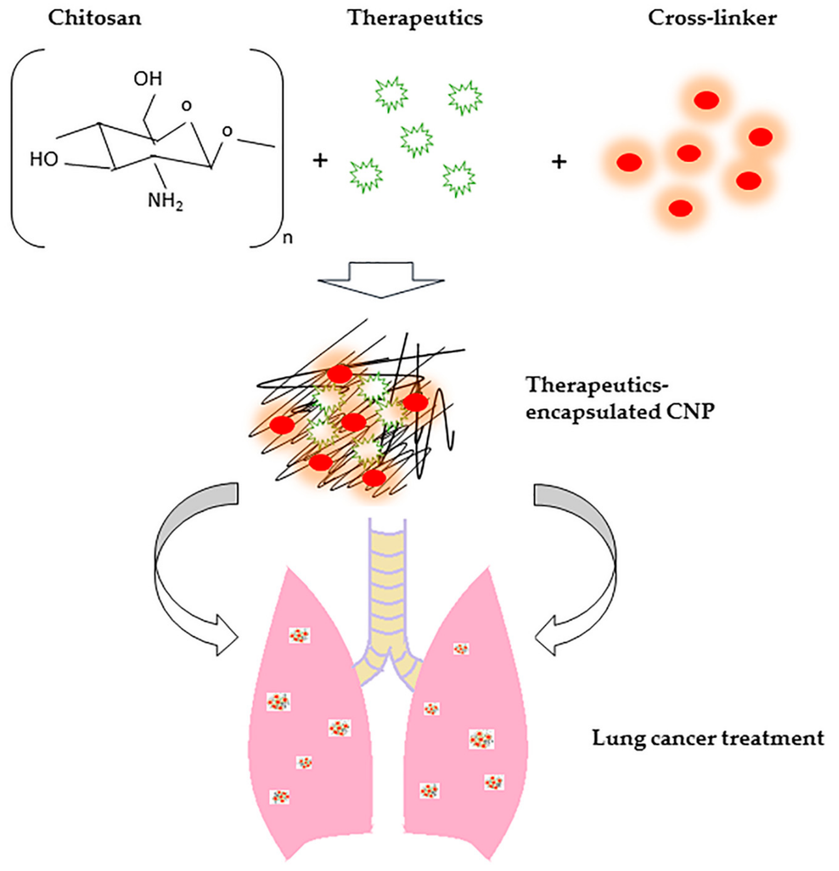
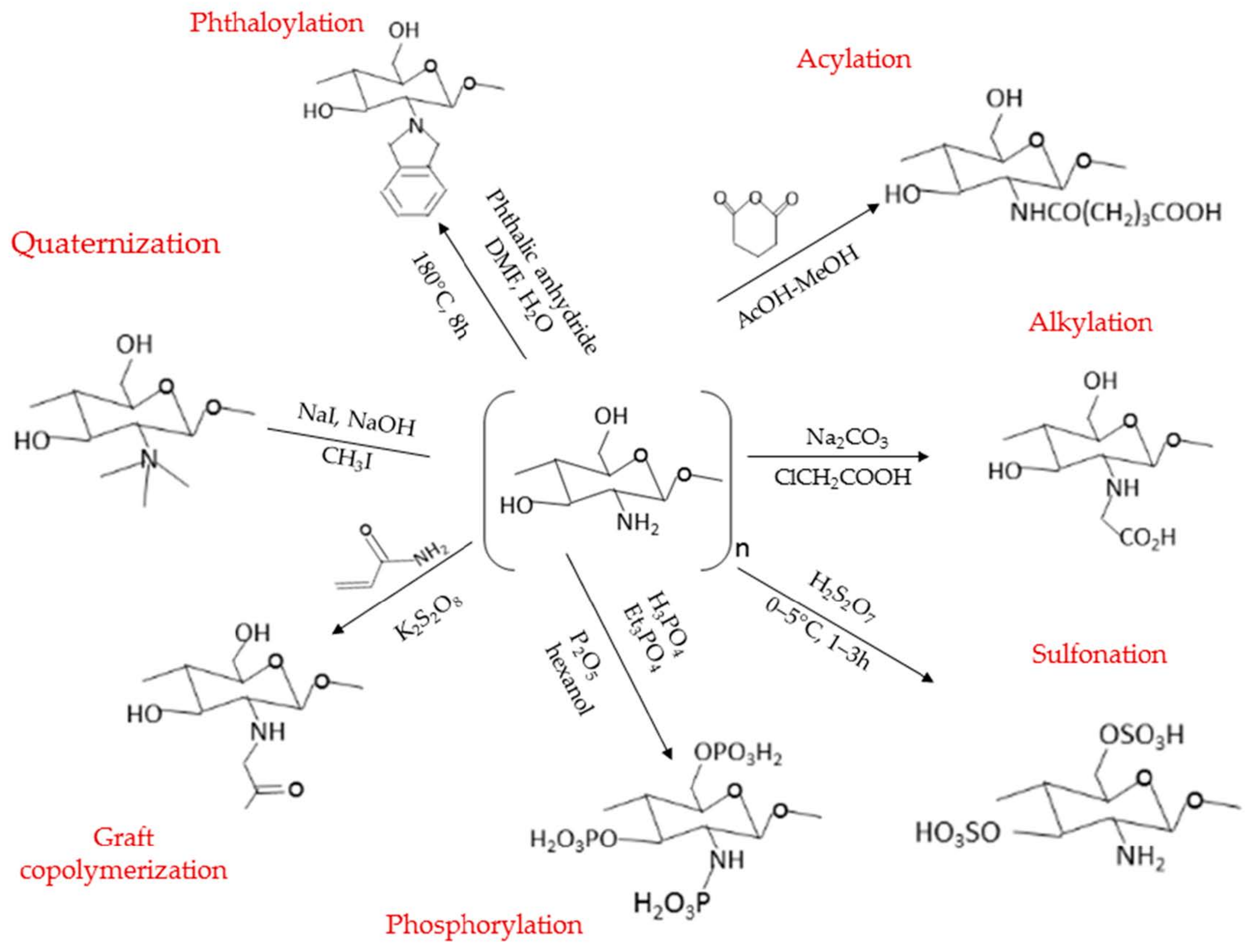
Table 1.
Applications of chemically-modified CNP-based systems.
| CS Derivatives | Application | Reference |
|---|---|---|
| Methylated N-Aryl CS Derivatives | An enhanced agent for in vitro paracellular permeation and in vivo adjuvant activity post oral administration to mice. | Suksamran et al. [185] |
| Quaternary CS magnetic composite modified with ammonium salt and combined with iron (II and III) oxide (Fe3O4) nanoparticles | A pH-dependent bioadsorption agent for methyl orange and chromium (VI) with homogeneous monolayer chemisorption behavior process. | Li et al. [186] |
| Acylated N,N,N-trimethyl CS nanopolyplexes associated with single-stranded oligonucleotides | Stable and nontoxic nanopolyplexes with enhanced cell transfection efficiency towards HeLa/Luc705 cell line. | Santos et al. [187] |
| N-acylated CS with glutaric anhydride in an aqueous acetic acid-methanol | Water soluble N-(4-carboxybutyroyl) CS derivative with improved antibacterial activity against Agrobacterium tumefaciens and Erwinia carotovora. and antifungal activity against Botrytis cinerea, Pythium debaryanum, and Rhizoctonia solani | Badawy and Rabea. [188] |
| O-(3,6-hydroxyethyl) CS | Improved water solubility, anticoagulation activity, and antibacterial activity against Escherichia coli. | Liu et al. [189] |
| Poly(ethylene glycol)-grafted-CS hydrogel | An improved delivery vector for delivery of T lymphocytes for brain tumor immunotherapy and potentially improved the glioblastoma immunotherapy. | Tsao et al. [190] |
| Polyurethane-grafted-CS copolymer | A biocompatible and hemocompatible copolymer with sustained release of tetracycline hydrochloride fitted with the Korsmeyer-Peppas release model. | Mahanta et al. [191] |
| 3,6-O-sulfonated CS | Human papillomavirus (HPV) infection inhibition via directly HPV capsids binding or indirectly blockage by host PI3K/Akt/mTOR pathway interference to prevent the entry of HPV16 through cell autophagy. | Gao et al. [192] |
| 2-hydroxyethylacrylate-grafted-CS | Controlled release of levofloxacin and proposed promising solution for topical wound management. | Siafaka et al. [193] |
| CS-N-2-hydroxypropyl trimethyl ammonium chloride | Enhanced water solubility and moisture-retention capacity, with antimicrobial activities against, Staphylococcus aureus, S. epidermidis, Bacillus subtilis, and Candida albicans. | Chi et al. [194] |
| Amino acid-grafted and N-acylated CS thiomers three-dimensional hydrogel scaffolds | A promising cytocompatible three-dimensional bio-scaffolds for potential cartilage repair applications. | Borsagli et al. [195] |
| Glutaraldehyde cross-linked carboxymethyl CS | Biocompatible with good hemostatic effect with an improved healing effect on liver injury in rats. | Zhang et al. [196] |
Table 2.
The outcome of drug-loaded CNP-based system for enhanced therapeutic efficacy/prolonged retention time/anti-MDR.
Table 2.
The outcome of drug-loaded CNP-based system for enhanced therapeutic efficacy/prolonged retention time/anti-MDR.
| CNP-Based System | Drug Loaded | Outcomes | Reference |
|---|---|---|---|
| Native CNP system | Curcumin (Cu) | In vitro studies revealed enhanced permeation of Cu through Strat-M® membrane and possessed controlled release properties in both pH 5.0 (first-order) and pH 7.4 (Higuchi) with negligible cytotoxicity towards human keratinocyte (HaCat) cell line compared to free Cu. | Nair et al. [56] |
| Native CNP system | Endostatin (ES) | The encapsulation of ES into CNP has prolonged the retention time of ES in vivo. The combined treatment of Lewis lung carcinoma (LLC) mouse Xenografts using ES-CNP and paclitaxel had significantly improved antitumor efficacy through suppressed proliferation and angiogenesis in the tumor tissues. | Xie et al. [246] |
| Native CNP system | Alphastatin (As) | In vitro studies have revealed high stability of the system and the sustained release properties of As for up to six days. Next, in vivo study of subcutaneous LA975 lung carcinoma xenograft in a T739 mouse model showed the greatest antiangiogenic effects and good hemocompatibility compared to the non-encapsulated counterparts. | Zhang and Hu [247] |
| Native CNP system | Docetaxel (Doc) | The in vitro studies showed the release of Doc from Doc-CNP using sustained-release proparticles (Higuchi release kinetic model) and possessed a significantly higher cytotoxicity effect in both dose- and time-dependent manners towards A549 cells compared to free Doc. The in vivo study using A549 xenograft nude mice also shown an enhanced anti-proliferative effect as compared with free Doc. | Nair and Velmurugan [248] |
| Folic acid conjugated CNP system (FLA-CNP) | Temozolomide (TMZ) | The TMZ-FLA-CNP was found to possess controlled- and sustained-release of TMZ with the highest antiproliferative efficacy against A549 lung cancer cell line as compared with TMZ-CNP and free TMZ. In vivo studies using A549 xenografted BALB/c-nu/nu athymic mice showed targeted delivery by pulmonary deposition and significantly higher tumor growth suppression as compared with TMZ-CNP and free TMZ. | Li et al. [249] |
| CS/ poly(lactic acid)/ graphene oxide/TiO2 composite nanofibrous scaffolds (CS/PLA/GO/TiO2) | Doxorubicin (Dox) | The CS/PLA/GO/TiO2/Dox showed the controlled-release of Dox (Korsmeyer–Peppas release kinetic model) for up to 14 days. The anti-proliferation efficacy of Dox was enhanced by the scaffolds and augmented with higher CS/PLA/TiO2/DOX/GO nanofibers concentration and the presence of magnetic field towards the A549 cell line. | Samadi et al. [250] |
| Carboxymethyl dextran conjugated CNP (CMD-CNP) | insulin-like growth factor 1 receptor specific siRNA (IGF-1R-siRNA) and doxorubicin (Dox) | The in vitro studies have presented the dual drug loading of IGF-1R-siRNA and Dox by CMD-CNP has enhanced the anti-migration, cytotoxicity, and apoptosis efficacy against A549 cell line as compared with a single-drug loading and free drug counterpart. | Shali et al. [251] |
| CS/poly(ethylene glycol)-anisamide (CTS/ PEG-AA) system | Gemcitabine (GEM) | The CTS/PEG-AA system showed the sustained-release of GEM for up to 15 days with enhanced in vitro cellular uptake and the most competitive cytotoxicity efficacy against A549 cells compared to CTS/PEG and free GEM. The in vivo study also showed superior tumor suppression against A549 subcutaneous tumors in mice in which revealed targeted delivery of GEM to the target site. | Garg et al. [252] |
| Native CNP system | Curcumin (Cu) | The Cu-CNP was released in a sustained-release manner for more than seven days and found with greater cytotoxicity efficacy even compared with Cu dissolved in DMSO solvent against HT1299 human lung cancer cell line. The in vivo studies using Swiss albino mice revealed that Cu-CNP possessed enhanced lung localization and were more competent in preventing benzo(a)pyrene-induced lung cancer as indicated by downregulation of proliferating cell nuclear antigen (PCNA), expression of p65 expression pERK. | Vijayakurup [253] |
| Nucleolin-targeting aptamer AS1411 and luminescent gold nanoclusters (AuNCs) functionalized CNP (AuNCs-CS-AS1411) system | Methotrexate (MTX) | MTX@AuNCs-CS-AS1411 showed the greatest in vitro anticancer effect against A549 cells compared to free MTX and MTX@AuNCs-CS as specified by an apoptotic death analysis. In vivo studies using A549 xenografted BALB/C nude mice have shown that AuNCs-CS-AS1411 conferred targeted delivery of MTX and effectively suppressed the tumor growth compared to free MTX. | Guo et al. [254] |
| Folate and carboxymethyl-β-cyclodextrin grafted trimethyl CNP system | Doxorubicin (Dox) and siRNA | Apart from showing satisfactory encapsulation efficiency, the grafted CNP system possessed pH-dependent controlled sustained release properties and significantly enhanced the therapeutic efficacy of the drugs, | Zhang et al. [255] |
| Cetuximab conjugated CNP (Cet-CNP) system | Quercetin (QUE) and Paclitacel (PTX) | The Que encapsulated Cet-CNP synergistically improved the cytotoxicity of PTX in A549 and reversed resistance in PTX resistant A549/Taxol cells. Besides, in vivo study revealed that the Que-PTX dual-loaded Cet-CNP had tumor growth inhibition in PTX-resistant xenografts. | Wang et al. [256] |
| Chitosan coated PLGA nanoparticles (CS-PLGA-NP) system | Resveratrol (RES) | The CS-PLGA-NP system showed improved stability and sustained release properties. Besides, the RES encapsulated CS-PLGA-NP showed significantly greater (about one-fold) cytotoxicity and apoptotic activities against H1299 lung cancer cells than the free drug counterpart. | Aldawsari et al. [257] |
| Native CNP system | Morinda citrifolia essential oil (MCEO) | The MCEOs-encapsulated CNPs had more than one-fold lower IC50 values as compared to free MCEOs (95 μg/mL, and 40 μg/mL, respectively) against A549 cells. | Rajivgandhi et al. [258] |
| Native CNP system | boswellic acid (BWA) | The BWA encapsulated CNP showed enhanced therapeutic effects against A549 cells because of greater cellular uptake, sustained-release properties, and enhanced antiproliferative effects as compared with free BWA. | Solanki et al. [259] |
| Hydrophobic deoxycholic acid (DCA) poly(amidoamine) dendronized CNP (DCA-PAMAM-CNP) system | Doxocubicin (Dox) and pDNA | The co-delivery of Dox and pDNA by using the DCA-PAMAM-CNP system achieved a high transfection efficiency of up to 74% in the 293T kidney cell line. Besides, a low dosage of co-delivered Dox was capable of improving transgene expression, presenting a synergistic effect. | Chen et al. [260] |
| Native CNP system | Anti-programmed cell death protein ligand 1 (aPD-L1) | The aerosol inhalation delivery system of aPD-L1 by CNP has been suggested to be a potent immunotherapy against lung metastasis through activation of the immune system by promoting the infiltration of different immune cells, especially CD8+ T cells. | Jin et al. [261] |
Publisher’s Note: MDPI stays neutral with regard to jurisdictional claims in published maps and institutional affiliations. |
© 2022 by the authors. Licensee MDPI, Basel, Switzerland. This article is an open access article distributed under the terms and conditions of the Creative Commons Attribution (CC BY) license (https://creativecommons.org/licenses/by/4.0/).
Share and Cite
MDPI and ACS Style
Yee Kuen, C.; Masarudin, M.J. Chitosan Nanoparticle-Based System: A New Insight into the Promising Controlled Release System for Lung Cancer Treatment. Molecules 2022, 27, 473. https://0-doi-org.brum.beds.ac.uk/10.3390/molecules27020473
AMA Style
Yee Kuen C, Masarudin MJ. Chitosan Nanoparticle-Based System: A New Insight into the Promising Controlled Release System for Lung Cancer Treatment. Molecules. 2022; 27(2):473. https://0-doi-org.brum.beds.ac.uk/10.3390/molecules27020473
Chicago/Turabian StyleYee Kuen, Cha, and Mas Jaffri Masarudin. 2022. "Chitosan Nanoparticle-Based System: A New Insight into the Promising Controlled Release System for Lung Cancer Treatment" Molecules 27, no. 2: 473. https://0-doi-org.brum.beds.ac.uk/10.3390/molecules27020473