Article

Flavonoid-Rich Fractions of Bauhinia holophylla Leaves Inhibit Candida albicans Biofilm Formation and Hyphae Growth

by
Sara Thamires Dias da Fonseca
1,
Thaiz Rodrigues Teixeira
2,
Jaqueline Maria Siqueira Ferreira
1,
Luciana Alves Rodrigues dos Santos Lima
1,
Walter Luyten
3 and
Ana Hortência Fonsêca Castro
1,*
1
Laboratory of Natural Products, Postgraduate Program in Biotechnology, Campus Centro-Oeste, Universidade Federal de São João del-Rei, Divinópolis 35501-296, MG, Brazil
2
Department of Biomolecular Sciences, School of Pharmaceutical Sciences of Ribeirão Preto, Universidade de São Paulo, Ribeirão Preto 14040-900, SP, Brazil
3
Department of Biology, Faculty of Science, Katholieke Universiteit Leuven, 3000 Leuven, Belgium
*
Author to whom correspondence should be addressed.
Submission received: 16 June 2022 / Revised: 29 June 2022 / Accepted: 29 June 2022 / Published: 7 July 2022

Abstract

:
This study evaluated the effect of the extract and fractions of Bauhinia holophylla on Candida albicans planktonic growth, biofilm formation, mature biofilm, and hyphae growth. Three C. albicans strains (SC5314, ATCC 18804, and ATCC 10231) were tested. The crude extract and the fractions were obtained by exhaustive percolation and liquid–liquid partition, respectively. Phytochemical analyses of B. holophylla extract and fractions were performed using high-performance liquid chromatography coupled with a diode-array detector and mass spectrometry (HPLC-DAD-MS). A microdilution assay was used to evaluate the effect of the B. holophylla extract and fractions on C. albicans planktonic growth, and crystal violet staining was used to measure the total biomass of the biofilm. Hyphae growth was analyzed using light microscopy. Thirteen flavonoids were identified, with a predominance of the flavonol-3-O-glycoside type based on quercetin, myricetin, and kaempferol. Flavonoid-rich fractions of B. holophylla leaves displayed antifungal activity and inhibited both biofilm formation and hyphae growth in all the tested strains, but were not effective on C. albicans planktonic growth and mature biofilm. This study indicates that flavonoid-rich fractions from B. holophylla leaves interfere with the virulence of Candida species and support the use of Bauhinia spp. in folk medicine to treat infections.

1. Introduction

Candida albicans is the major opportunistic pathogen of invasive fungal infections in humans [1,2]. Candida spp. infections represent 90% of invasive infections in hospitalized cases, mainly in patients undergoing intensive care, which can lead to death in up to 80% of cases [3,4]. Expression of adhesins and invasins on the cell surface, thigmotropism, the secretion of hydrolytic enzymes, the transition between yeast and hyphal forms, and biofilm formation are the main virulence factors of C. albicans [5]. The morphological transition between yeast and hyphal forms is an indicator of virulence because while the yeast form is identified as a disseminated form, the hyphal form is invasive to tissues and target cells and more resistant to phagocytosis, due to its ability to invade and kill macrophages [6,7].
In addition to the dimorphic (yeast–hyphae) transition, biofilm formation also increases virulence and fungal resistance by allowing Candida to grow and colonize inert surfaces, such as implants and urinary catheters, or host tissues, causing effects on human health [8,9]. Fungal biofilms are aggregates of microorganisms immobilized in a matrix containing extracellular polymeric substances of microbial origin [10,11]. Its formation occurs in four stages: (1) initial adhesion, (2) cell proliferation and the initial phase of filamentation, (3) biofilm maturation, and (4) release of yeast cells [12,13]. Candida cells found in a biofilm are more resistant to antimicrobials, UV radiation, dehydration, and disinfection, due to the complex three-dimensional architecture of the biofilm, the impermeable matrix, upregulation of efflux pumps, and metabolic plasticity [10,13,14]. Along with virulence factors, the emergence of strains resistant to available antifungal agents has increased due to new resistance mechanisms, and until now, there has been no specific antibiofilm drug [12]. Therefore, the treatment of fungal infections has become more difficult and burdens health systems across the world, requiring a search for new treatment alternatives.
Flavonoids are one of the major groups of bioactive compounds found in the Bauhinia genus. They correspond to an important class of plant-derived secondary metabolites, with demonstrated effects against bacteria, fungi, and viruses [15,16,17,18]. Several studies have reported the antibiofilm effects of flavonoids and their derivatives on bacteria and fungi. They are potential inhibitors of biofilm formation due to their ability to reduce the extracellular matrix [19,20], and they also inhibit the aggregation and maturation of the biofilm by decreasing surface hydrophobicity [21,22].
Bauhinia holophylla is a native species from the Brazilian Cerrado originally used in folk medicine to treat diabetes [23,24]. The hypoglycemic effects of the crude extracts of leaves were studied by Pinheiro et al. [24], Camaforte et al. [25], and Saldanha et al. [26]; in addition, Rozza et al. [27] observed their anti-ulcerogenic effects. Antiviral effects were also described by Santos et al. [28] against the dengue virus. Recently, Ribeiro et al. [29] and Marena et al. [30] reported the absence of cytotoxic and mutagenic effects, and even protective activity against known carcinogens, as well as antimicrobial effects of hydroalcoholic leaf extract, suggesting that B. holophylla has potential as a herbal medicine.
Currently, there are no reports on the antibiofilm activity of B. holophylla. Therefore, considering the presence of flavonoids in this species and the traditional use of some Bauhinia species to treat infections [31], this study aimed to investigate the antibiofilm activity of extracts and fractions of B. holophylla leaves against Candida albicans biofilm and hyphae growth.

2. Results

2.1. Phenolic Compounds’ Content

The total phenolic compounds’ contents in crude extract and fractions of B. holophylla leaves are shown in Table 1. The levels of total phenols and flavonoids ranged from 20.46 to 47.45 µg GAEq mg−1 DS and 2.37 to 9.93 µg QEq mg−1 DS, respectively. A higher total flavonoid content was observed in the DCM fraction, followed by the EtOAc fraction (p < 0.05).

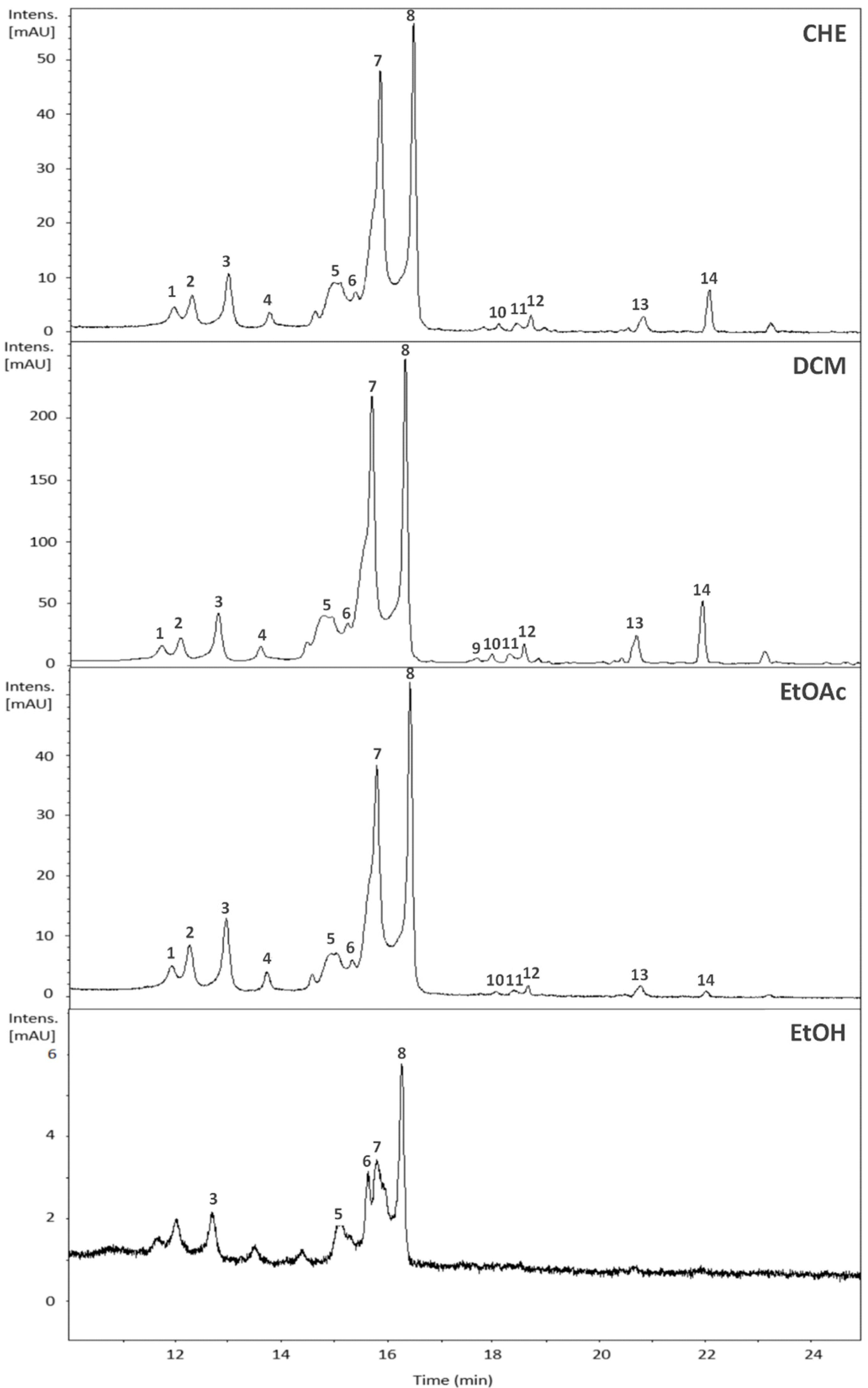
2.2. Chemical Composition of the Crude Extract and Fractions of B. holophylla

The samples of B. holophylla were analyzed using HPLC-DAD-MS to identify their chemical constituents (Figure 1, Table 2).
The patterns of fragmentation of the reference compounds reported in the literature, compared to those obtained in the present study, allowed us to identify 13 flavonoids, with predominance of the flavonol-3-O-glycoside type based on quercetin, myricetin, and kaempferol. The chromatogram peaks with absorption bands at 240-280 and 330-350 nm are typical of flavonoid derivatives of flavonols [32]. Mass fragments at m/z 303 and 319 characterized quercetin and myricetin, respectively. The ions at m/z [M+H]+ 435, 449, 465, and 479 were dereplicated as quercetin-O-pentoside, quercetin-O-deoxyhexoside, myricetin-O-hexoside, and myricetin-O-hexoside, respectively, similar to the results obtained by Camaforte et al. [25] and Rozza et al. [27]. The chemical profiles of the fractions are similar to that of the crude extract (CHE), except for the hydroethanolic (EtOH) fraction.

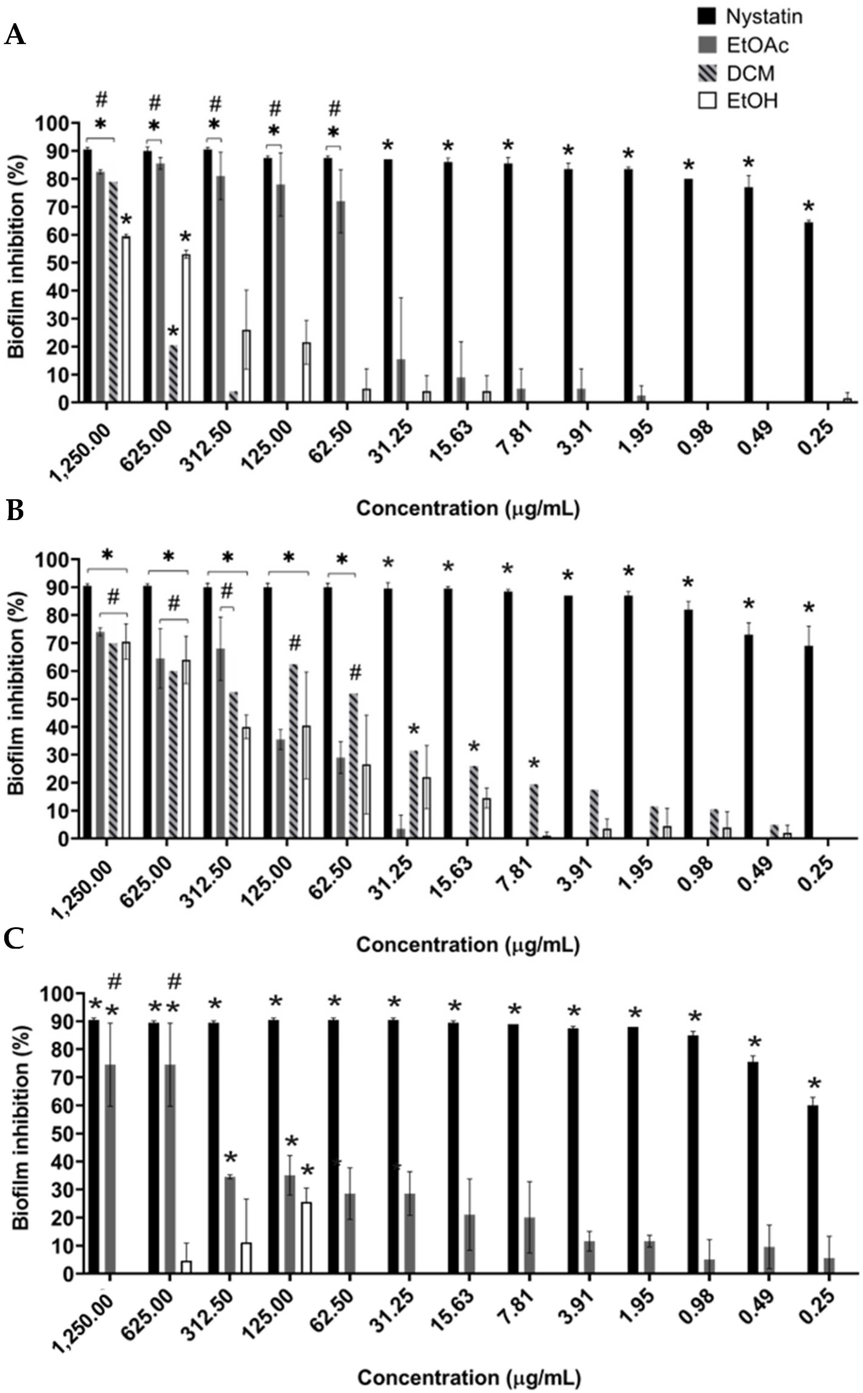
2.3. Evaluation of the Planktonic and Antibiofilm Activity against Candida albicans of Extract and Fractions

The planktonic growth of C. albicans strains was not altered when treated with the B. holophylla extract and fractions for 48 h (Table 3). However, C. albicans strains were susceptible to the positive controls, as expected. C. albicans SC5314 and C. albicans ATCC 18804 growth decreased significantly due to fluconazole (MIC= 1.95 µg/mL) and. C. albicans ATCC 10231 due to nystatin (MIC= 3.91 µg/mL).

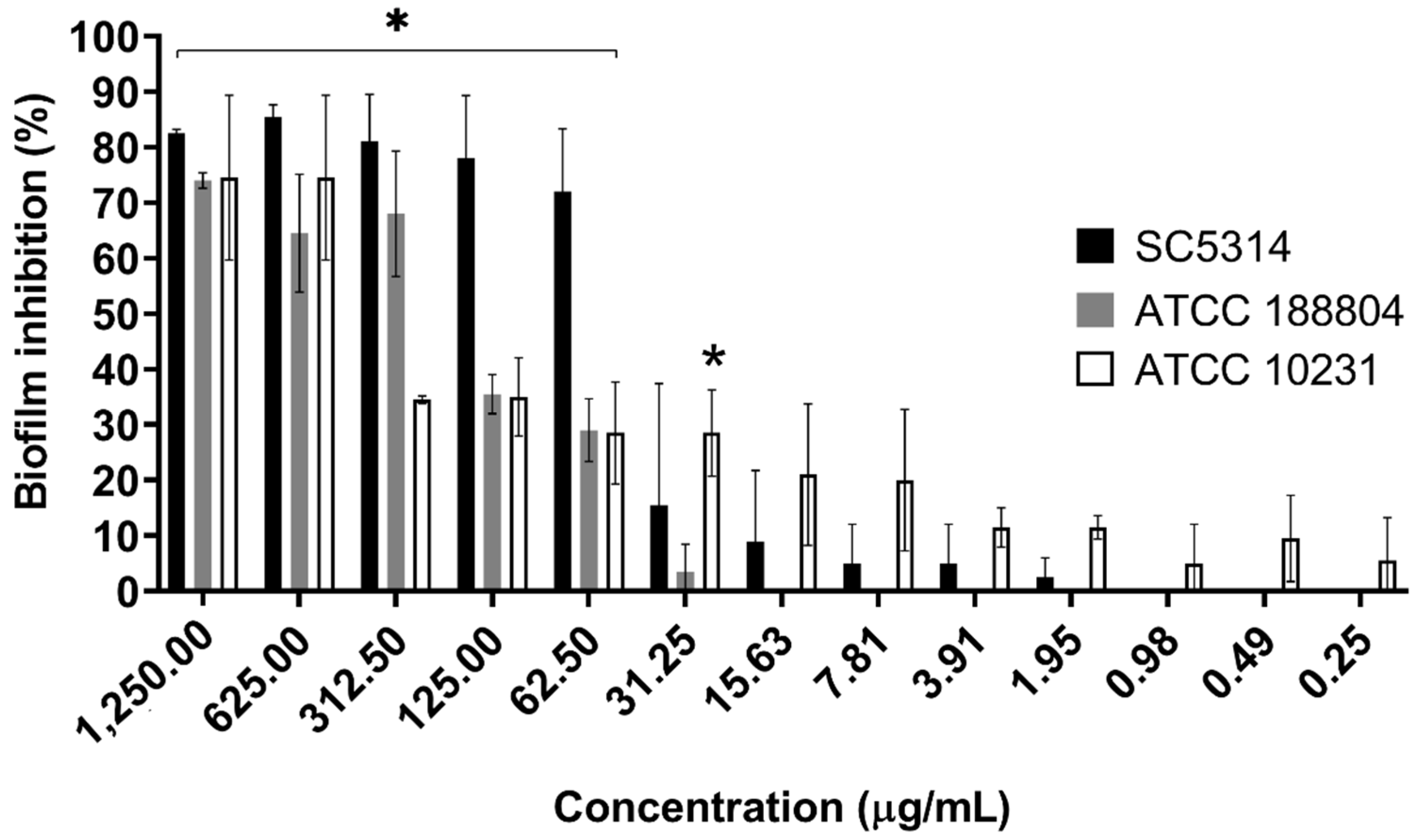
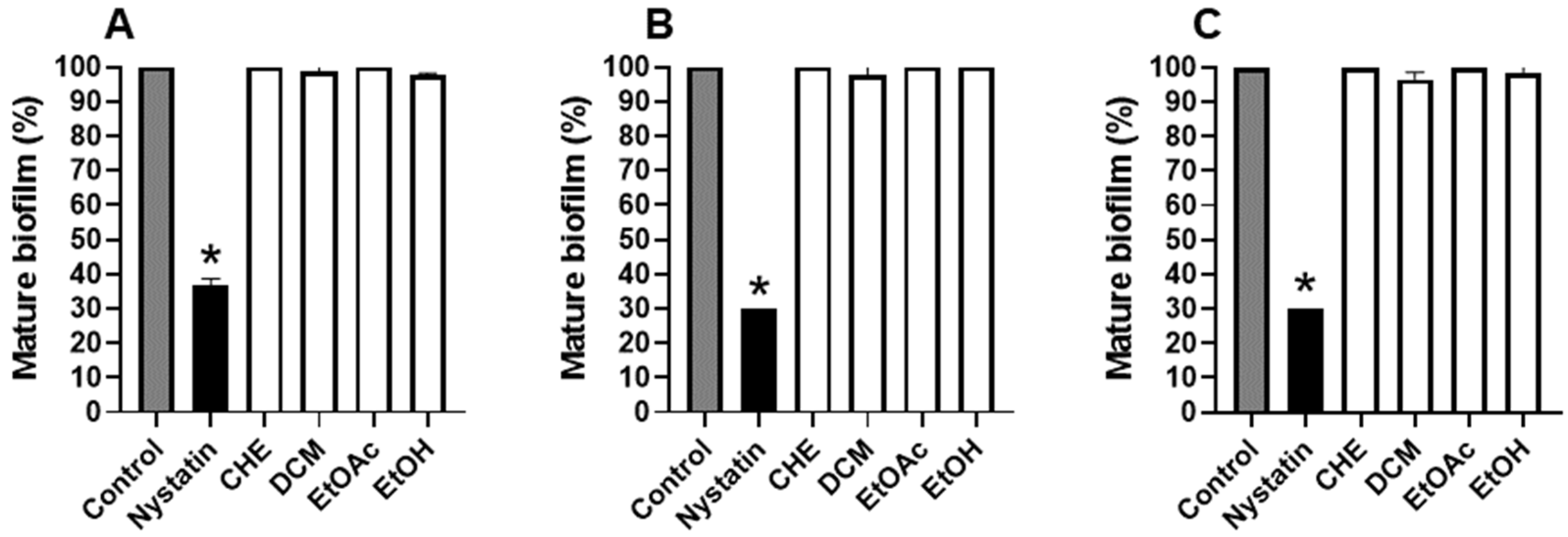
2.4. B. holophylla Fractions Inhibit Biofilm Formation of C. albicans Strains, but Do Not Have Effect on Mature Biofilm

C. albicans biofilm formation was not inhibited by B. holophylla extract, but the EtOAc, DCM, and EtOH fractions were effective in all of the tested strains. The three fractions tested decreased C. albicans SC5314 and C. albicans ATCC 18804 biofilms, and the EtOAc and EtOH fractions showed inhibitory effects against C. albicans ATCC 10231 (p < 0.05). Biofilm formation by C. albicans strains was dramatically inhibited by nystatin, the positive control (Figure 2).
C. albicans SC5314 biofilm formation decreased significantly with the EtOAc fraction with a highly inhibitory effect from 1250 to 62.5 μg/mL (82 to 72% inhibition) (Figure 2A). An inhibitory effect similar to the nystatin (about 90%) was observed at a concentration of 125 μg/mL, and from 31.25 μg/mL, the inhibitory effect dropped to 18% and was progressively reduced further in the other concentrations (p < 0.05). C. albicans SC5314 biofilm formation was also affected by DCM fraction, especially at 1250 μg/mL with 79% inhibition and an EtOH fraction at 1250 to 625 μg/mL (60 and 55% inhibition, respectively). B. holophylla fractions significantly inhibited C. albicans ATCC 18804 biofilm formation (p < 0.05) (Figure 2B). Inhibition was greater than 60% at 1250 to 625 μg/mL for all the fractions. EtOAc and DCM maintained this inhibition at 312.5 and 125 μg/mL, respectively. From 312.5 μg/mL, a progressive reduction in the inhibitory effect of the EtOH fraction was observed. C. albicans ATCC 10231 biofilm formation decreased significantly in a concentration-dependent manner with the EtOAc fraction (p < 0.05) (Figure 2C). The maximum inhibitory effect (75%) was reached when it was treated with 1250 and 625 μg/mL of the EtOAc fraction. From 312.5 to 7.8 μg/mL, the inhibitory effect decreased to about 35 to 20%, and from 3.9 to 0.25 μg/mL, the inhibition was approximately 10% on average.
This result was less prominent for the EtOH fraction, which showed some inhibition at concentrations from 625 to 125 µg/mL, with 27% inhibition of biofilm formation at 125 µg/mL.
Results show a potential inhibition of C. albicans biofilm formation by B. holophylla fractions, especially EtOAc, which presented a marked inhibition for all three strains tested (Figure 3). This effect was more pronounced on the SC5314 strain, where the inhibition of biofilm formation was greater than 80% at concentrations between 1250 and 312.5 μg/mL, and remained above 70% for concentrations from 125 to 62.5 μg/mL.
Although the fractions had a pronounced inhibitory effect on biofilm formation, extract and fractions of B. holophylla did not have an effect on mature the biofilm of C. albicans strains (p > 0.05) (Figure 4).

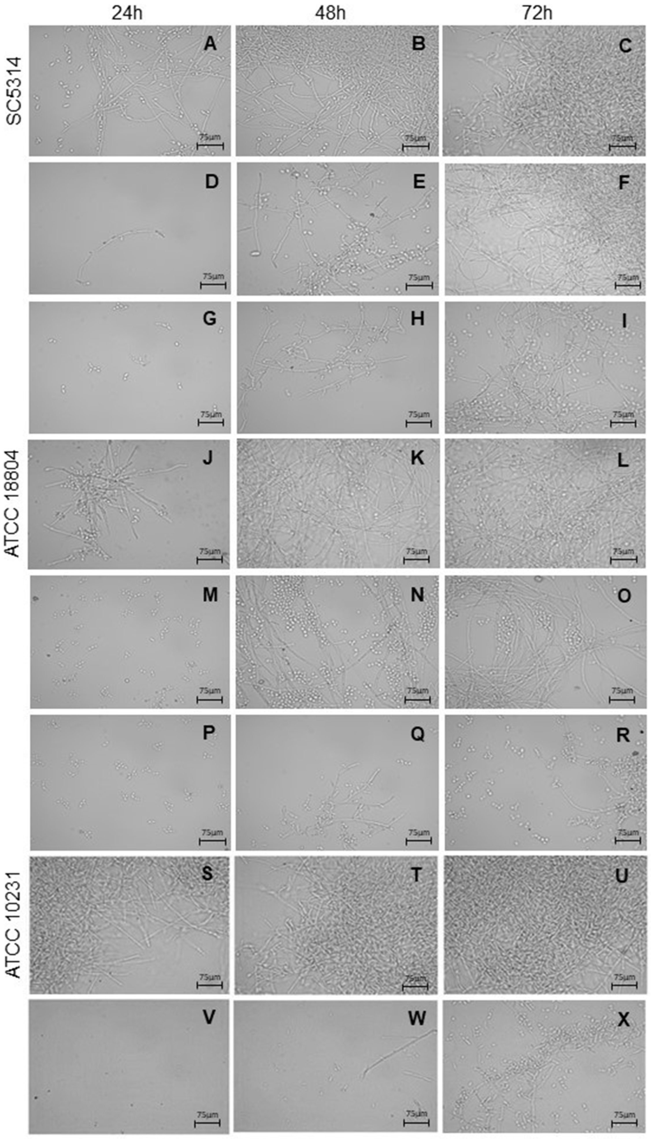
2.5. B. holophylla Fractions Reduce Yeast–Hyphae Transition

The fractions of B. holophylla were able to reduce the yeast–hyphae transition at 625 µg/mL, compared to untreated cells, depending on the studied strain (Figure 5). These effects could be observed mainly after 24 and 48 h of incubation. The B. holophylla DCM fraction reduced the yeast–hyphae transition of C. albicans SC5314, since growth was reduced at 24 and 48 h (Figure 5A–I). Treatment of C. albicans ATCC 18804 with the B. holophylla EtOAc fraction markedly reduced the yeast–hyphae transition at 24, 48, and 72 h, while the DCM fraction was active mainly in the first 24 h (Figure 5J–R). For C. albicans ATCC 10231, the EtOH fraction led to the lowest number of cells growing in the hyphal form after 24, 48, and 72 h of incubation (Figure 5S–X). The yeast–hyphae transition of C. albicans strains was dramatically inhibited by nystatin, the positive control.

3. Discussion

The therapeutic effects of several medicinal plants are attributed to the abundant presence of flavonoids as bioactive compounds [33]. Flavonoids are ubiquitous natural products found in the plant kingdom, often in a glycoside form. Flavonols correspond to one of the main classes of flavonoids, being chemically a 3-hydroxy derivative of flavones [9,33]. Their wide distribution in nature, low cost, and low toxicity are advantages that make flavonoids good candidates for application in antifungal therapy [34].
In the current study, we observed that fractions of B. holophylla leaves containing, predominantly, flavonoids of the flavonol-3-O-glycoside type were able to inhibit biofilm formation of C. albicans strains and the yeast–hyphae transition in vitro. However, these fractions showed no effect on C. albicans planktonic growth and mature biofilms. The ethyl acetate fractions, containing flavonol-3-O-glycosides based on quercetin, myricetin, and kaempferol, exhibited a marked activity against biofilm formation on all strains tested, but especially on the SC5314 strain. Similarly, these fractions were able to reduce the yeast–hyphae transition at 625 µg/mL, depending on the studied strain.
Bauhinia spp. are traditionally used in folk medicine to treat infections, pain, inflammations, and diabetes [31]. Several of these activities have been attributed to flavonoids and scientifically proven by in vivo and in vitro models [25,28,35]. Currently, there are few studies on the antimicrobial effects of B. holophylla. Marena et al. [30] reported antibacterial and antifungal effects against some microorganisms, but these activities have not been confirmed by Fonseca et al. [36]. The reason for this discrepancy is not clear.
Bioguided studies of extracts and fractions of B. holophylla leaves have led to the identification of several glycosylated flavonoids and aglycones [25,27]. There are complex glycosylated flavonoids with up to five sugar residues, and some studies have shown that different glycosylated flavonoids exert different biological activities [37]. Flavonoids can be synthesized in plants in response to microbial infection. However, the mechanisms of antifungal activity and the antivirulence properties of flavonoids remain unknown [33,38,39].
B. holophylla extract and fractions did not inhibit C. albicans growth, but they were able to inhibit biofilm formation and also reduced the yeast–hyphae transition. Most C. albicans infections are associated with biofilm formation, an important virulence factor, since the biofilm is resistant to most available antifungal drugs [40]. In addition, the dimorphic transition from a budding yeast cell to a filamentous form (yeast–hyphae) also represents another important virulence factor, since the hyphae contribute to the stability of biofilms, and their ability to adhere is critical for tissue penetration [12,13,41]. These factors not only facilitate adherence to and penetration into the host tissue, but they also evade host immunity [42]. These two virulence attributes are primary sources of multiple-drug-resistance development and invasive infections, because they are difficult or even impossible to eradicate with conventional antifungal agents [43,44]. In this way, the flavonoid-rich fractions of B. holophylla leaves are potential candidates for use in combination with established antifungal drugs to treat C. albicans infections, since they may act in different stages of biofilm formation. However, some studies showed that antibiofilm activity can vary among Bauhinia spp., since flavonoids from a B. forficata extract exhibited strong antifungal activity at 15.62 µg/mL on the planktonic form, but lower inhibition on biofilm formation (45%) [45]. Therefore, preventing biofilm and hyphae formation could facilitate the treatment of C. albicans infections, because the number of available anticandidal drugs classes is limited [46].
Flavonoids of the flavonol-3-O-glycoside type are an important group of chemical compounds that also have a broad-spectrum antifungal activity. Quercetin, myricetin, kaempferol, and their glycosylated forms have been reported as therapeutic molecules for human health, and emerge as promising novel antifungals [47,48,49]. Despite their antifungal activity, some reports demonstrate that when applied alone, quercetin, e.g., exhibits low antifungal activity, but in combined therapy with fluconazole, there is a strong synergism in the clinical management of C. albicans biofilms [44,50]. Just like quercetin, myricetin also has multiple biological actions [51], and some studies showed that myricetin has antifungal and potent antibiofilm activities, therefore being able to enhance the antifungal effect of miconazole in combined therapy [52]. Quercetin and myricetin also have potential to control hyphae growth [53]. In addition, kaempferol, an active flavonoid, has been considered a potential candidate drug against Candida species, and the concomitant use of fluconazole and kaempferol demonstrated a kaempferol-induced reversion in fluconazole-resistant C. albicans [46].

4. Materials and Methods

4.1. Chemicals

The following chemicals were purchased from different manufacturers: nystatin (Pharma Nostra, Rio de Janeiro, Brazil), fluconazole (Fagron, São Paulo, Brazil), ethanol (AlphaTec®, Santo André, Brazil), hexane, dichloromethane, and ethyl acetate (Cromato Produtos Químicos®, Diadema, Brazil), Sabouraud Dextrose Broth (Acumedia, San Bernardino, CA, USA), and Fetal Bovine Serum (Gibco®, São Paulo, Brazil). Dimethylsulphoxide (DMSO), crystal violet, RPMI 1640 (Roswell Park Memorial Institute Medium, Buffalo, New York), and MOPS (3-[N-Morpholino] propane sulfonic acid) were purchased from Sigma-Aldrich Co. (St. Louis, MI, USA). RPMI1640 medium (without NaHCO3 and with L-glutamine) was buffered with 0.165 M of MOPS to pH 7.4.

4.2. Plant Material and Preparation of the Extract and Fractions

Bauhinia holophylla (Bong.) Steud. (Fabaceae: Cercideae) leaves were collected in the Brazilian Cerrado in Ijaci, Southern Minas Gerais State, Brazil (21°09′97″ S and 44°55′65″ W GRW, at 835 m altitude) (SISBIO n° 24542-3, IBAMA Registration: 5042260). Fertile samples were collected, and the vouchers were identified by Andreia Fonseca Silva of the PAMG Herbarium (PAMG 57021) at the Agricultural Research Company of Minas Gerais (EPAMIG). This work has access permission for plant genetic heritage components (No. 010500/2014-6/CNPq/CGEN/MMA), and is registered on the SisGen Platform (Register A12A940), according to the Brazilian Biodiversity Law (13.123/2015).
The plant material was dried in a ventilated oven (TE-394/500L, Tecnal; Piracicaba, Brazil) at 40 °C for 24 h, and pulverized in a knife mill (SL-31, Solab; Piracicaba, Brazil). The dried and powdered leaves (400 g) were extracted by exhaustive percolation using 70% ethanol as the extraction solvent for 7 days. The solvent was removed on a rotary evaporator (R-220PRO, Büchi do Brazil; Valinhos, Brazil) at 50 °C under reduced pressure, obtaining the crude hydroethanolic extract (CHE, 46.60 g; yield = 11.65%). The CHE (5 g) was solubilized in 70% ethanol (200 mL) and subsequently subjected to liquid–liquid partition in a separating funnel with n-hexane, dichloromethane, and ethyl acetate. The solvents were removed using a rotary evaporator (R-220PRO, Büchi do Brazil; Valinhos, Brazil) at 50°C, under reduced pressure, resulting in dichloromethane (DCM, 0.926 g), ethyl acetate (EtOAc, 0.945 g), and hydroethanolic (EtOH, 0.964 g) fractions. The yield of the n-hexane fraction was negligable.

4.3. Phenolic Compounds Content

Total phenols were quantified using the Folin–Ciocalteau method according to Pastrana-Bonilla et al. [54]. The total phenol content was calculated using a calibration curve with 100 µg mL−1 gallic acid solution as the standard. Sample absorbances were read at 760 nm using a UV–visible spectrophotometer (Q798Ul, Quimis; Diadema, Brazil). Determinations were performed in triplicate, and the results are given in microgram equivalents of gallic acid per milligram of dry sample (µg GAEq mg−1 DS).
The total flavonoid assay was performed according to Woisky and Salatino [55], and flavonoid content was calculated using a calibration curve with 100 µg mL−1 quercetin in a methanol solution of 2% aluminum chloride as a standard. Sample absorbances were read at 425 nm using a UV–visible spectrophotometer (Q798U, Quimis; Diadema, Brazil). Determinations were performed in triplicate, and the results are given in microgram equivalents of quercetin per milligram of dry sample (µg QEq mg−1 DS).

4.4. HPLC-DAD-MS Analyses

The chemical composition of the B. holophylla crude extract and its fractions were analyzed on an ultra-fast liquid chromatography (UFLC) system (Prominence, Shimadzu; Kyoto, Japan), using an Ascentis Express C18 column (10 cm × 4.6 mm, 2.7 µm (Supelco Analytical; Bellefonte, PA, USA). This was coupled to a diode-array detector (DAD) (SPD-M20A) monitored between 200 and 800 nm, and a quadrupole time-of-flight tandem mass spectrometer (micrOTOF QII, Bruker Daltonics; Massachusetts, USA) equipped with an electrospray ionization (ESI) source. The injection volume was 1 μL of the sample (prepared at a concentration of 1 mg/mL), and the flow rate was 0.3 mL/min. The mobile phase was composed of acetonitrile (solvent B) and deionized water (solvent A) with the addition of 0.1% formic acid (v/v). The applied elution profile was: 0–5 min: 10 to 20% B, 5–10 min: 20% B, 10–15 min: 20 to 30% B, 15–20 min: 30 to 45% B, 20–33 min: 45 to 100% B, 33–37 min: 100% B, 37–38 min: 100 to 10% B, and 38–42 min: 10% B. Mass spectrometry analysis was applied using the following parameters: capillary voltage, 3.5 kV; m/z range, 120–1300; source and desolvation temperature, 220 °C; cone and desolvation gas flow rate, 9.0 L/min. Nitrogen (4 Bar) was used as the collision gas for MS/MS analyses, with collision energies ranging from 25 to 62.5 eV. The high-resolution mass spectrometer was calibrated using a TFA-Na+ solution (10 mg/mL). Identification of the compounds in the samples was attained by comparing the retention times, UV, and MS spectra with literature data.

4.5. Antibiofilm Formation Assay

4.5.1. Strains and Culture Conditions

Three Candida albicans strains, including C. albicans SC5314 (ATCC MYA-2876), C. albicans ATCC 18804, and C. albicans ATCC 10231, were employed in this study. All the strains were originally obtained from the American Type Culture Collection (ATCC). C. albicans SC5314 and C. albicans ATCC 18804 were kindly provided by Dr. Susana Johann (Microbiology Laboratory, Federal University of Minas Gerais). According to ATCC (2015), C. albicans ATCC SC5314 and C. albicans ATCC 18804 are sensitive to anidulafungin, voriconazole, itraconazole, fluconazole, micafungin, caspofungin, and 5-flucytosine. C. albicans ATCC 10231 is resistant to anidulafungin, voriconazole, itraconazole, fluconazole, and ketoconazole, but is sensitive to micafungin, caspofungin, and 5-flucytosine. The fungal strain cultures were routinely maintained in incubation at 37 °C (SP-101/30, SPLabor; Presidente Prudente, Brazil) in 2% Sabouraud dextrose broth [56].

4.5.2. Microdilution Assay: Effect of the B. holophylla Extract and Fractions on C. albicans Planktonic Growth

The microdilution method was employed to determine the susceptibility of C. albicans strains in planktonic growth according to document M27-A3 of the Clinical and Laboratory Standards Institute, with minor modifications [57]. The B. holophylla extract and fractions were freshly dissolved in 2% DMSO at a concentration of 2.5 mg/mL and then two-fold diluted serially to the desired concentrations. In this study, 1 × 10³ CFU/mL of the C. albicans strains was treated with B. holophylla extract and fractions (0.25–1250 µg/mL) by the addition of the respective samples and C. albicans suspension in Sabouraud dextrose broth into 96-well round-bottom microplates. Fluconazole was used as a positive control for C. albicans SC5314 and C. albicans ATCC 18804, and nystatin was used as a positive control for C. albicans ATCC 10231. Cells treated with 2% (v/v) DMSO were used as the vehicle control. Samples under each condition were incubated at 37 °C for 48 h, and the minimum inhibitory concentration (MIC) was determined as the lowest concentration where no visible growth was observed. The assays were performed in triplicate, and the results are expressed as the mean of the three independent experiments.

4.5.3. Antibiofilm Formation Assay

Biofilm formation was determined as described previously by Xu et al. [58], with minor modifications. For the adhesion stage, C. albicans strains (100 μL) were seeded at a density of 1 × 10⁶ CFU/mL in 96-well flat-bottom microplates, and incubated at 37 °C for 90 min to allow attachment of the yeast cells on the polystyrene surface. After this, non-adherent cells were removed, and adhered cells were cultured in the presence or absence of B. holophylla extract or fractions, freshly dissolved in 2% DMSO at a concentration of 0.25–1250.00 µg/mL. Fresh RPMI 1640 medium (100 µL/well) was added, and the plates were incubated at 37 °C for 48 h.
To study the effect of B. holophylla extract or fractions on mature biofilm, yeast cells were suspended in RPMI 1640 medium (1 × 10⁶ CFU/mL), and 100 µL was transferred into the wells of flat-bottom 96-well plates, and incubated at 37 °C for 48 h to allow biofilm formation and maturation. Next, the biofilms were treated with the extract or fractions (1250 µg/mL) for 48 h. Crystal violet staining (0.1%) was used to measure total biomass [59]. Data are expressed as percentages of biofilm mass in treated samples vs. untreated controls. Nystatin was used as positive control, and RPMI 1640 medium supplemented with 2% (v/v) DMSO was used as the vehicle control.

4.5.4. Inhibition of Yeast–Hyphae Transition

The effect of B. holophylla extract or fractions on yeast–hyphae transition was evaluated according to Andrade et al. [60]. Hyphae growth was induced by incubation of C. albicans strains (1 × 10³ CFU/mL) in a 96-well microplate with fetal bovine serum and B. holophylla extract or fractions (625, 312.50, and 125 µg/mL). The microplates were incubated for 24, 48, and 72 h at 37 °C, and hyphae formation was observed with a light microscope (Primo Star, Zeiss; São Paulo, Brazil) using 400-fold magnification and documented by AxioVision software (Zeiss). The experiments were performed in triplicate, and repeated three times. Positive (nystatin) and solvent (2% v/v DMSO) controls were included.

4.6. Statistical Analysis

All tests were performed in triplicate with at least two independent experiments. The software used for the statistics was GraphPad Prism® v. 8.0.1 (GraphPad Software, Inc. La Jolla; California, USA). An one-way analysis of variance (ANOVA) test was used, followed by a Tukey’s test to compare the results between groups, and Dunnet’s test was used to compare the results between the treatments and the control, with a p-value < 0.05 deemed significant. The results are expressed as the mean ± standard deviation.

5. Conclusions

The qualitative analysis of the chemical profile of extract and fractions from B. holophylla leaves revealed the presence of flavonoids, predominantly of the flavonol-3-O-glycoside type based on quercetin, myricetin, and kaempferol. The flavonoid-rich fractions showed a promising effect on biofilm formation and hyphae growth, important virulence factors of C. albicans strains. The results confirm the biotechnological potential of Bauhinia spp. to produce bioactive compounds of economic and medicinal interest, and suggest that further investigations aimed at the isolation and biological evaluation of isolated compounds are desirable.

Author Contributions

Conceptualization, Walter Luyten and A.H.F.C.; Data curation, S.T.D.d.F. and T.R.T.; Formal analysis, S.T.D.d.F. and T.R.T.; Funding acquisition, J.M.S.F., L.A.R.d.S.L. and A.H.F.C.; Investigation, S.T.D.d.F., J.M.S.F., L.A.R.d.S.L. and A.H.F.C.; Methodology, S.T.D.d.F. and T.R.T.; Project administration, A.H.F.C.; Resources, J.M.S.F., L.A.R.d.S.L. and A.H.F.C.; Supervision, A.H.F.C.; Writing—original draft, S.T.D.d.F., T.R.T. and A.H.F.C.; Writing—review and editing, T.R.T., J.M.S.F., W.L. and A.H.F.C. All authors have read and agreed to the published version of the manuscript.

Funding

Conselho Nacional de Desenvolvimento Científico e Tecnológico (CNPq) (Processo 444907/2014-9), and Fundação de Amparo à Pesquisa do Estado de Minas Gerais (FAPEMIG) (CRA-APQ-01347-14). This study was financed in part by the Coordenação de Aperfeiçoamento de Pessoal de Nível Superior—Brasil (CAPES), Finance Code 001.

Data Availability Statement

The data presented in this study are available within the article.

Acknowledgments

We thank Andreia Fonseca Silva for the botanical identification of this plant, and Hosana Maria Debonsi for authorization to carry out the HPLC analysis by T.R.Teixeira. The authors are grateful to the Universidade Federal de São João del-Rei (UFSJ), Conselho Nacional de Desenvolvimento Científico e Tecnológico (CNPq), Coordenação de Aperfeiçoamento de Pessoal de Nível Superior (CAPES), and Fundação de Amparo à Pesquisa do Estado de Minas Gerais (FAPEMIG) for financial support.

Conflicts of Interest

The authors declare no conflict of interest.

References

  1. Singla, R.K.; Dubey, A.K. Molecules and metabolites from natural products as inhibitors of biofilm in Candida spp. pathogens. Curr. Top. Med. Chem. 2019, 19, 2567–2578. [Google Scholar] [CrossRef] [PubMed]
  2. Wall, G.; Montelongo-Jauregui, D.; Bonifacio, B.V.; Lopez-Ribot, J.L.; Uppuluri, P. Candida albicans biofilm growth and dispersal: Contributions to pathogenesis. Curr. Opin. Microbiol. 2019, 52, 1–6. [Google Scholar] [CrossRef] [PubMed]
  3. Dadar, M.; Tiwari, R.; Karthik, K.; Chakraborty, S.; Shahali, Y.; Dhama, K. Candida albicans-biology, molecular characterization, pathogenicity, and advances in diagnosis and control—An update. Microb. Pathog. 2018, 117, 128–138. [Google Scholar] [CrossRef]
  4. Pristov, K.E.; Ghannoum, M.A. Resistance of Candida to azoles and echinocandins worldwide. Clin. Microbiol. Infect. 2019, 25, 792–798. [Google Scholar] [CrossRef] [PubMed]
  5. Mayer, F.L.; Wilson, D.; Hube, B. Candida albicans pathogenicity mechanisms. Virulence 2013, 15, 119–128. [Google Scholar] [CrossRef] [Green Version]
  6. Sudbery, P.; Gow, N.; Berman, J. The distinct morphogenic states of Candida albicans. Trends Microbiol. 2004, 12, 317–324. [Google Scholar] [CrossRef] [PubMed]
  7. Correia, I.; Román, E.; Prieto, D.; Hidalgo-Vico, S.; Alonso-Monge, R.; Pla, J. Role of Candida albicans mating in genetic variability and adaptation to the host. Fungal Biol. Rev. 2019, 33, 180–189. [Google Scholar] [CrossRef]
  8. Stacy, A.; Mcnally, L.; Darch, S.E.; Brown, S.P.; Whiteley, M. The biogeography of polymicrobial infection. Nat. Rev. Microbiol. 2016, 14, 93–105. [Google Scholar] [CrossRef] [Green Version]
  9. Guzzo, F.; Scognamiglio, M.; Fiorentino, A.; Buommino, E.; D’Abrosca, B. Plant derived natural products against Pseudomonas aeruginosa and Staphylococcus aureus: Antibiofilm activity and molecular mechanisms. Molecules 2020, 25, 5024. [Google Scholar] [CrossRef]
  10. Aguiar, F.L.L.; Santos, N.C.; de Paula Cavalcante, C.S.; Andreu, D.; Baptista, G.R.; Gonçalves, S. Antibiofilm activity on Candida albicans and mechanism of action on biomembrane models of the antimicrobial peptide Ctn [15–34]. Int. J. Mol. Sci. 2020, 21, 8339. [Google Scholar] [CrossRef]
  11. D’Acunto, B.; Frunzo, L.; Klapper, I.; Mattei, M.R.; Stoodley, P. Mathematical modeling of dispersal phenomenon in biofilms. Math. Biosci. 2019, 307, 70–87. [Google Scholar] [CrossRef] [PubMed] [Green Version]
  12. Gulati, M.; Nobile, C.J. Candida albicans biofilms: Development, regulation, and molecular mechanisms. Microbes Infect. 2016, 18, 310–321. [Google Scholar] [CrossRef] [PubMed] [Green Version]
  13. Höfs, S.; Mogavero, S.; Hube, B. Interaction of Candida albicans with host cells: Virulence factors, host defense, escape strategies, and the microbiota. J. Microbiol. 2016, 54, 149–169. [Google Scholar] [CrossRef]
  14. Cavalheiro, M.; Teixeira, M.C. Candida biofilms: Threats, challenges, and promising strategies. Front. Med. 2018, 5, 28. [Google Scholar] [CrossRef] [PubMed] [Green Version]
  15. Farag, M.A.; Sakna, S.T.; El-Fiky, N.M.; Shabana, M.M.; Wessjohann, L.A. Phytochemical, antioxidant and antidiabetic evaluation of eight Bauhinia L. species from Egypt using UHPLC–PDA–qTOF-MS and chemometrics. Phytochemistry 2015, 119, 41–50. [Google Scholar] [CrossRef] [PubMed]
  16. Farhadi, F.; Khameneh, B.; Iranshahi, M.; Iranshahy, M. Antibacterial activity of flavonoids and their structure-activity relationship: An uptade review. Phytother. Res. 2018, 33, 13–40. [Google Scholar] [CrossRef] [Green Version]
  17. Jin, Y. Recent advances in natural antifungal flavonoids and their derivatives. Bioorg. Med. Chem. Lett. 2019, 29, 126589. [Google Scholar] [CrossRef]
  18. Ninfali, P.; Antonelli, A.; Magnani, M.; Scarpa, E.S. Antiviral properties of flavonoids and delivery strategies. Nutrients 2020, 12, 2534. [Google Scholar] [CrossRef]
  19. Matilla-Cuenca, L.; Gil, C.; Cuesta, S.; Rapún-Araiz, B.; Ziemyté, M.; Mira, A.; Lasa, I.; Vale, J. Antibiofilm activity of flavonoids on staphylococcal biofilms through targeting BAP amyloids. Sci. Rep. 2020, 10, 18968. [Google Scholar] [CrossRef]
  20. Pruteanu, M.; Lobato, J.I.H.; Stach, T.; Hengge, R. Common plant flavonoids prevent the assembly of amyloid curli fibres and can interfere with bacterial biofilm formation. Environ. Microbiol. 2020, 22, 5280–5299. [Google Scholar] [CrossRef]
  21. Kanchanapiboon, J.; Kongsa, U.; Pattamadilok, D.; Kamponchaidet, S.; Wachisunthon, D.; Poonsatha, S.; Tuntoaw, S. Boesenbergia rotunda extract inhibits Candida albicans biofilm formation by pinostrobin and pinocembrin. J. Ethnopharmacol. 2020, 261, 113193. [Google Scholar] [CrossRef] [PubMed]
  22. Tao, J.; Yan, S.; Zhou, C.; Liu, Q.; Zhu, H.; Wen, Z. Total flavonoids from Potentilla kleiniana wight et arn inhibits biofilm formation and virulence factors production in methicillin-resistant Staphylococcus aureus (MRSA). J. Ethnopharmacol. 2021, 279, 114383. [Google Scholar] [CrossRef] [PubMed]
  23. Vaz, A.M.S.F.; Bortulozzi, R.L.C.; da Silva, L.A.E. Checklist of Bauhinia sensu stricto (Caesalpiniaceae) in Brazil. Plant Ecol. Evol. 2010, 143, 212–221. [Google Scholar] [CrossRef]
  24. Pinheiro, M.S.; Rodrigues, L.S.; Neto, L.S.; Moraes-Souza, R.Q.; Soares, T.S.; Americo, M.F. Effect of Bauhinia holophylla treatment in streptozotocin-induced diabetic rats. An. Acad. Bras. Cienc. 2017, 89, 263–272. [Google Scholar] [CrossRef]
  25. Camaforte, N.A.D.P.; Saldanha, L.L.; Vareda, P.M.P.; Rezende-Neto, J.M.; Senger, M.R.; Delgado, A.Q.; Morgan, H.J.N.; Violato, N.M.; Pieroni, L.G.; Dokkedal, A.L.; et al. Hypoglycaemic activity of Bauhinia holophylla through GSK3-β inhibition and glycogenesis activation. Pharm. Biol. 2019, 57, 269–279. [Google Scholar] [CrossRef] [Green Version]
  26. Saldanha, L.L.; Delgado, A.Q.; Marcourt, L.; Camaforte, N.A.P.; Vareda, P.M.P.; Ebrahimi, S.N.; Vilegas, W.; Dokkedal, A.L.; Queiroz, E.F.; Wolfender, J.; et al. Hypoglycemic active principles from the leaves of Bauhinia holophylla: Comprehensive phytochemical characterization and in vivo activity profile. PLoS ONE 2021, 16, e0258016. [Google Scholar] [CrossRef]
  27. Rozza, A.L.; Cesar, D.A.S.; Pieroni, L.G.; Saldanha, L.L.; Dokkedal, A.L.; De-Faria, F.M.; Souza-Brito, A.R.M.; Vilegas, W.; Takahira, R.K.; Pellizzon, C.H. Antiulcerogenic activity and toxicity of Bauhinia holophylla hydroalcoholic extract. Evid. Based Complement. Altern. Med. 2015, 2015, 439506. [Google Scholar] [CrossRef]
  28. dos Santos, M.; Teixeira, T.R.; Santos, F.R.S.; Lima, W.G.; Ferraz, A.C.; Silva, N.L.; Leite, F.J.; Siqueira, J.M.; Luyten, W.; Castro, A.H.F.; et al. Bauhinia holophylla (Bong.) Steud. leaves-derived extracts as potent anti-dengue serotype 2. Nat. Prod. Res. 2021, 35, 2804–2809. [Google Scholar] [CrossRef]
  29. Ribeiro, D.L.; Cilião, H.L.; Specian, A.F.L.; Serpeloni, J.M.; de Oliveira, M.T.; Varanda, E.A.; Vilegas, W.; Saldanha, L.L.; Martínez-López, W.; Dokkedal, A.L.; et al. Phytochemical study and evaluation of cytotoxicity, mutagenicity, cell cycle kinetics and gene expression of Bauhinia holophylla (Bong.) Steud. in HepG2 cells in vitro. Cytotechnology 2018, 70, 713–728. [Google Scholar] [CrossRef] [Green Version]
  30. Marena, G.D.; Saldanha, L.L.; Ramos, M.A.S.; de Grandis, R.A.; Dokkedal, A.L.; Bauab, T.M.; Pavan, F.R.; Resende, F.A. Antimicrobial, cytotoxic and mutagenic activities of Bauhinia holophylla hydroalcoholic extract. Am. J. Essent. Oil. Nat. Prod. 2021, 9, 1–6. [Google Scholar]
  31. Filho, V.C. Chemical Composition and biological potential of plants from the genus Bauhinia. Phytother. Res. 2009, 23, 1347–1354. [Google Scholar] [CrossRef] [PubMed]
  32. Andersen, O.M.; Markham, K.R. Flavonoids: Chemistry, Biochemistry and Applications, 1st ed.; CRC Press: Boca Raton, FL, USA, 2006; pp. 617–856. ISBN 0-8493-2021-6. [Google Scholar]
  33. Ivanov, M.; Kannan, A.; Stojkovic, D.S.; Glamoclija, J.; Calhelha, R.C.; Ferreira, I.C.F.R.; Sanglard, D.; Sokovic, M. Flavones, flavonols, and glycosylated derivatives-impact on Candida albicans growth and virulence, expression of CDR1 and ERG11, cytotoxicity. Pharmaceuticals 2020, 14, 27. [Google Scholar] [CrossRef]
  34. Ansari, M.A.; Fatima, Z.; Hameed, S. Sesamol: A natural phenolic compound with promising anticandidal potential. J. Pathog. 2014, 2014, 895193. [Google Scholar] [CrossRef] [PubMed] [Green Version]
  35. Miceli, N.; Buongiorno, L.P.; Celi, M.G.; Cacciola, F.; Dugo, P.; Donato, P.; Mondello, L.; Bonaccorsi, I.; Taviano, M.F. Role of the flavonoid-rich fraction in the antioxidant and cytotoxic activities of Bauhinia forficata Link. (Fabaceae) leaves extract. Nat. Prod. Res. 2015, 30, 1229–1239. [Google Scholar] [CrossRef] [PubMed]
  36. Fonseca, S.T.D.; Teixeira, T.R.; Ferreira, J.M.S.; Lima, L.A.R.S.; Siqueira, J.M.; Luyten, W.; Castro, A.H.F. Phenolic compounds and antioxidant and antibacterial activities of Bauhinia holophylla (Fabaceae:Cercideae). In Phenolic Compounds in Health and Disease, 1st ed.; Linn, R.C., Ed.; Nova Science Publishers: New York, NY, USA, 2021; pp. 153–173. ISBN 978-1-68507-137-0. [Google Scholar]
  37. Rochefort, S.J.; Imsic, M.; Jones, R.; Trenerry, V.C.; Thomkins, B. Characterization of flavonol conjugates in immature leaves of pak choi [Brassica rapa L. sp. chinensis L. (Hanelt.)] by HPLC- DAD and LC-MS/MS. J. Agric. Food. Chem. 2006, 54, 4655–4860. [Google Scholar] [CrossRef]
  38. Mishra, B.; Leishangthem, G.D.; Gill, K.; Singh, A.K.; Das, S.; Singh, K.; Xess, I.; Dinda, A.; Kapil, A.; Patro, I.K.; et al. A novel antimicrobial peptide derived from modified N-terminal domain of bovine lactoferrin: Design, synthesis, activity against multidrug-resistant bacteria and Candida. Biochim. Biophys. Acta. Biomembr. 2013, 1828, 677–686. [Google Scholar] [CrossRef] [Green Version]
  39. Smiljković, M.; Kostić, M.; Stojković, D.; Glamočlija, J.; Soković, M. Could flavonoids compete with synthetic azoles in diminishing Candida albicans infections? A comparative review based on in vitro studies. Curr. Med. Chem. 2019, 26, 2536–2554. [Google Scholar] [CrossRef]
  40. Wall, G.; Chaturvedi, A.K.; Wormley, F.L.; Wiederhold, N.P.; Patterson, H.P.; Patterson, T.F.; Lopez-Ribot, J.L. Screening a repurposing library for inhibitors of multidrug-resistant Candida auris identifies ebselen as a repositionable candidate for antifungal drug development. Antimicrob. Agents Chemother. 2018, 62, e01084-18. [Google Scholar] [CrossRef] [PubMed] [Green Version]
  41. Nobile, C.J.; Johnson, A.D. Candida albicans biofilms and human disease. Annu. Ver. Microbiol. 2015, 69, 71–92. [Google Scholar] [CrossRef] [Green Version]
  42. Patel, M.; Srivastava, V.; Ahmad, A. Dodonaea viscosa var angustifolia derived 5,6,8-trihydroxy-7,4′dimethoxy flavone inhibits ergosterol synthesis and the production of hyphae and biofilm in Candida albicans. J. Ethnopharmacol. 2020, 259, 112965. [Google Scholar] [CrossRef]
  43. Lewis, K. Persister cells, dormancy and infectious disease. Nat. Ver. Microbiol. 2007, 5, 48–56. [Google Scholar] [CrossRef] [PubMed]
  44. Singh, B.N.; Upreti, D.K.; Singh, B.R.; Pandey, G.; Verma, S.; Roy, S.; Naqvi, A.H.; Rawat, A.K.S. Quercetin sensitizes fluconazole-resistant Candida albicans to induce apoptotic cell death by modulating quorum sensing. Antimicrob. Agents Chemother. 2015, 59, 2153–2168. [Google Scholar] [CrossRef] [PubMed] [Green Version]
  45. Alves, E.P.; Lima, R.F.; de Almeida, C.M.; Freires, I.A.; Rosalen, P.L.; Ruiz, A.L.; Granville-Garcia, A.F.; Godoy, G.P.; Pereira, J.V.; Costa, E.M.M.B. Antimicrobial and antiproliferative activity of Bauhinia forficata Link and Cnidoscolus quercifolius extracts commonly used in folk medicine. J. Contemp. Dent. Pract. 2017, 18, 635–640. [Google Scholar] [CrossRef] [PubMed]
  46. Shao, J.; Zhang, M.; Wang, T.; Li, Y.; Wang, C. The roles of CDR1, CDR2, and MDR1 in kaempferol-induced suppression with fluconazole-resistant Candida albicans. Pharm. Biol. 2016, 54, 984–992. [Google Scholar] [CrossRef] [PubMed] [Green Version]
  47. Semwal, D.; Semwal, R.; Combrinck, S.; Viljoen, A. Myricetin: A dietary molecule with diverse biological activities. Nutrients 2016, 8, 90. [Google Scholar] [CrossRef] [Green Version]
  48. Imran, M.; Rauf, A.; Shah, Z.A.; Saeed, F.; Imran, A.; Arshad, M.U.; Ahmad, B.; Bawazeer, S.; Atif, M.; Peters, D.G.; et al. Chemo-preventive and therapeutic effect of the dietary flavonoid kaempferol: A comprehensive review. Phytother. Res. 2018, 33, 263–275. [Google Scholar] [CrossRef]
  49. Salehi, B.; Machin, L.; Monzote, L.; Sharifi-Rad, J.; Ezzat, S.M.; Salem, M.A.; Merghany, R.M.; Mahdy, N.M.E.; Kiliç, C.S.; Sytar, O.; et al. Therapeutic potential of quercetin: New insights and perspectives for human health. ACS Omega 2020, 5, 11849–11872. [Google Scholar] [CrossRef]
  50. Gao, M.; Wang, H.; Zhu, L. Quercetin assists fluconazole to inhibit biofilm formations of fluconazole-resistant Candida albicans in in vitro and in vivo antifungal managements of vulvovaginal candidiasis. Cell. Physiol. Biochem. 2016, 40, 727–742. [Google Scholar] [CrossRef]
  51. Song, X.; Tan, L.; Wang, M.; Ren, C.; Guo, C.; Yang, B.; Ren, Y.; Cao, Z.; Li, Y.; Pei, J. Myricetin: A review of the most recent research. Biomed. Pharmacother. 2021, 134, 111017. [Google Scholar] [CrossRef]
  52. Mo, F.; Ma, J.; Yang, X.; Zhang, P.; Li, Q.; Zhang, J. In vitro and in vivo effects of the combination of myricetin and miconazole nitrate incorporated to thermosensitive hydrogels, on C. albicans biofilms. Phytomedicine 2020, 71, 153223. [Google Scholar] [CrossRef]
  53. Mishra, S.; Rastogi, S.K.; Singh, S.; Panwar, S.L.; Shrivash, M.K.; Misra, K. Controlling pathogenesis in Candida albicans by targeting EFG1 and glyoxylate pathway through naturally occurring polyphenols. Mol. Biol. Rep. 2019, 46, 5805–5820. [Google Scholar] [CrossRef] [PubMed]
  54. Pastrana-Bonilla, E.; Akoh, C.C.; Sellappan, S.; Krewer, G. Phenolic content and antioxidant capacity of muscadine grapes. J. Agric. Food Chem. 2003, 51, 5497–5503. [Google Scholar] [CrossRef] [PubMed]
  55. Woisky, R.G.; Salatino, A. Analysis of propolis: Some parameters and procedures for chemical quality control. J. Apic. Res. 1998, 37, 99–105. [Google Scholar] [CrossRef]
  56. Clinical and Laboratory Standarts Institute (CLSI). Methods for Dilution Antimicrobial Susceptibility Tests for Bacteria that Grow Aerobically, 90th ed.; Clinical and Laboratory Standards Institute: Wayne, NJ, USA, 2012; CLSI, Document M07-A9. [Google Scholar]
  57. Clinical and Laboratory Standarts Institute (CLSI). Reference Method for Broth Dilution Antifungal Susceptibility Testing of Yeasts, 3rd ed.; Clinical and Laboratory Standards Institute: Wayne, NJ, USA, 2008; CLSI, Document M27-A3. [Google Scholar]
  58. Xu, K.; Wang, J.L.; Chu, M.P.; Jia, C. Activity of coumarin against Candida albicans biofilms. J. Mycol. Med. 2018, 29, 28–34. [Google Scholar] [CrossRef] [PubMed]
  59. Liu, N.; Zhang, N.; Zhang, S.; Zhang, L.; Liu, Q. Phloretin inhibited the pathogenicity and virulence factors against Candida albicans. Bioengineered 2021, 12, 2420–2431. [Google Scholar] [CrossRef] [PubMed]
  60. Andrade, J.T.; Santos, F.R.S.; Lima, W.G.; Sousa, C.D.F.; Oliveira, L.S.F.M.; Ribeiro, R.I.M.A.; Gomes, A.J.P.S.; Araújo, M.G.F.; Villar, J.A.F.P.; Ferreira, J.M.S. Design, synthesis, biological activity and structure-activity relationship studies of chalcone derivatives as potential anti-Candida agentes. J. Antibiot. 2018, 71, 702–712. [Google Scholar] [CrossRef]
Figure 1. Expansion of total ion chromatograms (10–25 min) of CHE (crude hydroethanolic extract), DCM (dichloromethane fraction), EtOAc (ethyl acetate fraction), and EtOH (hydroethanolic fraction) from B. holophylla.
Plants 11 01796 g001
Figure 2. Effect of Bauhinia holophylla fractions on biofilm formation of Candida albicans strains. (A) C. albicans SC5314; (B) C. albicans ATCC 18804; (C) C. albicans ATCC 10231. The metabolic activity represented by the C. albicans biofilm was compared with the untreated control. The results between groups were compared using Tukey’s test, and Dunnett’s test was used to compare the results between the treatments and the control. Each value is presented as the mean ± standard deviation of two independent experiments (* p-value < 0.05). (#) fractions and concentrations do not differ from each other. EtOAc: ethyl acetate fraction; DCM: dichloromethane fraction; EtOH: hydroethanolic fraction.
Plants 11 01796 g002
Figure 3. Reduction in biofilm formation of C. albicans strains by ethyl acetate fraction. The control represents the culture without any treatment, defined as 0% inhibition. Each concentration had seven replicates. The results between groups were compared using Tukey’s test, and Dunnet’s test was used to compare the results between the treatments and the control. Each value is presented as the mean ± standard deviation of two independent experiments (* p-value < 0.05).
Plants 11 01796 g003
Figure 4. Effects of the Bauhinia holophylla extract and fractions (1250 µg/mL) on mature biofilm of C. albicans. (A) C. albicans SC5314; (B) C. albicans ATCC 18804; (C) C. albicans ATCC 10231. The control is the absence of treatment and 100% biofilm biomass. The results between groups were compared with Tukey’s test, and Dunnet’s test was used to compare the results between the treatments and the control. The results are presented as the mean ± standard deviation of two independent experiments (* p-value < 0.05). CHE: crude hydroethanolic extract; DCM: dichloromethane fraction; EtOAc: ethyl acetate fraction; EtOH: hydroethanolic fraction.
Plants 11 01796 g004
Figure 5. Hyphal formation by Candida albicans strains. C. albicans was cultured in the absence (control) and presence of Bauhinia holophylla extract and fractions at 625, 312.5, and 125 µg/mL for 24, 48, and 72 h at 37 °C. Nystatin was used as a positive control. Only active samples are shown at 625 µg/mL. (AC) C. albicans SC5314 control; (DF) SC5314 with crude hydroethanolic extract; (GI) SC5314 with dichloromethane fraction; (JL) C. albicans ATCC 18804 control; (MO) ATCC 18804 with dichloromethane fraction; (PR) ATCC 18804 with ethyl acetate fraction; (SU) C. albicans ATCC 10231 control; (VX) ATCC 10231 with hydroethanolic fraction. Representative microphotographs were obtained using 400-fold magnification. Scale bars: 75 µM.
Plants 11 01796 g005
Table 1. Total phenol and flavonoid contents in the extract and fractions of B. holophylla leaves. CHE: crude hydroethanolic extract; DCM: dichloromethane fraction; EtOAc: ethyl acetate fraction; EtOH: hydroethanolic fraction. The results were compared using Tukey’s test, and they are presented as the mean ± standard deviation of three repetitions. Means with the same letter do not differ statistically from each other (p-value < 0.05).
SampleTotal Phenols
(µg GAEq mg−1 DS)
Total Flavonoids
(µg QEq mg−1 DS)
CHE39.08 ± 1.77 b3.91 ± 0.13 c
DCM37.61 ± 2.54 b9.93 ± 0.53 a
EtOAc47.45 ± 1.51 a6.03 ± 0.35 b
EtOH20.46 ± 2.32 c2.37 ± 0.13 c
[Reprinted from: Book, Phenolic Compounds in Health and Disease, Phenolic Compounds and Antioxidant and Antibacterial Activities of Bauhinia holophylla (Fabaceae: Cercideae), 153–174, Copyright: 2021, Nova Science Publishers, Inc., Authors: Sara Dias da Fonseca, Thaiz Rodrigues Teixeira, Jaqueline Maria Siqueira Ferreira, Luciana Alves Rodrigues dos Santos Lima, João Máximo de Siqueira, Walter Luyten, Ana Hortência Fonseca Castro].
Table 2. Characterization of compounds in crude extract and fractions from Bauhinia holophylla using high-performance liquid chromatography coupled with diode array detector and mass spectrometry (HPLC-DAD-MS) in positive ionization mode. CHE (crude hydroethanolic extract); EtOAc (ethyl acetate fraction); DCM (dichloromethane fraction); EtOH (hydroethanolic fraction). (+) presence and (-) absence. NI = not identified.
PeakRtUVmax [M + H]+ (m/z)[M + H]+ (m/z)ErrorMS/MSnCompoundSamplesMolecular Formula
(min.)(nm)ExperimentalTheoretical(ppm)CHEDCMEtOAcEtOH
112.0254/350451.0855451.08713.5319.0412; 303.0472Myricetin-O-pentoside+++-C20H18O12
212.3255/348465.1013465.10283.2319.0441; 303.0511Myricetin-O-deoxyhexoside+++-C21H20O12
313.0254/353465.1004465.10285.2303.0496Quercetin-O-hexoside++++C21H20O12
413.8254/350465.1024465.10280.9303.0503Quercetin-O-hexoside+++-C21H20O12
515.0255/350435.0900435.09225.0303.0493Quercetin-O-xilopyranose++++C20H18O11
615.6255/349449.1062449.10783.6287.0559Kaempferol-3-O-glucoside++++C21H20O11
715.9255/348435.0901435.09224.8303.0499Quercetin-O-pentoside++++C20H18O11
816.5254/348449.1056449.10784.9303.0470Quercetin-O-deoxyhexoside++++C21H20O11
917.7264/346419.0959419.09733.3287.0505Kaempferol-O-pentoside-+--C20H18O10
1018.1264/346479.1168479.11843.3317.0624; 302.0381Isorhamnetin-3-O-hexoside+++-C22H22O12
1118.4264/348479.0938--317.0619Undentified+++-NI
1218.7264/345433.1108433.11294.8287.0517Luteolin-deoxyhexose+++-C21H20O10
1320.8254/362303.0493303.04992.0181.9561Quercetin+++-C15H10O7
1422.0254/356317.0645317.06563.5302.0408Isorhamnetin+++-C16H12O7
Table 3. Minimal inhibitory concentration (MIC) of extract and fractions of Bauhinia holophylla against Candida albicans strains. CHE: crude hydroethanolic extract; DCM: dichloromethane fraction; EtOAc: ethyl acetate fraction; EtOH: hydroethanolic fraction; NYS: nystatin; FLU: fluconazole. (-) not tested. The results were compared using Dunnet’s test, and they are presented as the mean of three repetitions. Means with the same letter do not differ statistically from each other (p-value < 0.05).
Candida albicansMIC (µg/mL)Drugs
CHEDCMEtOAcEtOHFLUNYS
SC5314>1250 c>1250 c>1250 c>1250 c1.95 a-
ATCC 18804>1250 c>1250 c>1250 c>1250 c1.95 a-
ATCC 10231>1250 c>1250 c>1250 c>1250 c-3.91 b
Publisher’s Note: MDPI stays neutral with regard to jurisdictional claims in published maps and institutional affiliations.

