Article

Suppressive Effects of Rosmarinic Acid Rich Fraction from Perilla on Oxidative Stress, Inflammation and Metastasis Ability in A549 Cells Exposed to PM via C-Jun, P-65-Nf-Κb and Akt Signaling Pathways

by
Komsak Pintha
1,
Wittaya Chaiwangyen
1,
Supachai Yodkeeree
2,
Maitree Suttajit
1 and
Payungsak Tantipaiboonwong
1,*
1
Division of Biochemistry, School of Medical Sciences, University of Phayao, Phayao 56000, Thailand
2
Department of Biochemistry, Faculty of Medicine, Chiang Mai University, Chiang Mai 50200, Thailand
*
Author to whom correspondence should be addressed.
Submission received: 9 July 2021 / Revised: 19 July 2021 / Accepted: 20 July 2021 / Published: 23 July 2021

Abstract

:
Particulate matter from forest fires (PMFF) is an environmental pollutant causing oxidative stress, inflammation, and cancer cell metastasis due to the presence of polycyclic aromatic hydrocarbons (PAHs). Perilla seed meal contains high levels of polyphenols, including rosmarinic acid (RA). The aim of this study is to determine the anti-oxidative stress, anti-inflammation, and anti-metastasis actions of rosmarinic acid rich fraction (RA-RF) from perilla seed meal and its underlying molecular mechanisms in A549 cells exposed to PMFF. PMFF samples were collected via the air sampler at the University of Phayao, Thailand, and their PAH content were analyzed using GC-MS. Fifteen PAH compounds were detected in PMFF. The PMFF significantly induced intracellular reactive oxygen species (ROS) production, the mRNA expression of pro-inflammatory cytokines, MMP-9 activity, invasion, migration, the overexpression of c-Jun and p-65-NF-κB, and Akt phosphorylation. Additionally, the RA-RF significantly reduced ROS production, IL-6, IL-8, TNF-α, and COX-2. RA-RF could also suppress MMP-9 activity, migration, invasion, and the phosphorylation activity of c-Jun, p-65-NF-κB, and Akt. Our findings revealed that RA-RF has antioxidant, anti-inflammatory, and anti-metastasis properties via c-Jun, p-65-NF-κB, and Akt signaling pathways. RA-RF may be further developed as an inhalation agent for the prevention of lung inflammation and cancer metastasis induced by PM exposure.

1. Introduction

In the northern region of Thailand, air pollution is an annual and severe environmental issue. Long-term exposure to air pollution causes both acute and chronic health effects [1]. The burning of the forests and agricultural wastes are associated with smoke pollution in this region [2]. Fine particulate matter ≤10 μm (PM10, PM2.5) has been identified as one of the most prevalent human health risks due to its deep infiltration into the respiratory tract [3,4]. These particles have the capacity to accumulate various toxic components, including polycyclic aromatic hydrocarbons (PAHs) [5]. PAHs carry potential mutagens and carcinogens that can promote inflammation and tumor progression [6,7]. PM can activate free radical species generation, which strongly influences the development of oxidative DNA damage, eventually causing DNA mutation [6].
Unbalanced reactive oxygen species (ROS) production and antioxidant activity increases oxidative stress that can trigger a cascade of inflammation signaling pathways and cancer cell metastasis [8,9]. PM-induced oxidative stress has been accepted as a key molecular mechanism of many inflammatory modulators, such as tumor necrosis factor-α (TNF-α), interleukins IL-1, IL-6, IL-8, and COX-2, including MMP-9 expression and cancer cell metastasis [10,11,12,13]. The elevation of inflammatory mediators and the MMP-9 degradation enzyme are associated with Akt, NF-KB, and AP1 signaling pathways [14].
Previous studies have demonstrated that polyphenol extract from medicinal plants suppressed ROS production, proinflammatory cytokine expression, and MMP degradation enzymes in PM-treated cells [15,16]. Perilla frutescens L. (Nga-mon in Thai), belonging to the mint family, has been widely used for consumption and as a medicinal herb in Asian countries including Thailand, Japan, Korea, China, and India [17]. Chemically, the perilla seed contains high levels of essential fatty acids, mainly α-linolenic acid (~60%), linoleic acid (~20%) [18], and phenolic compounds. Among these phenolic acids, rosmarinic acid (RA) is reportedly predominant in the ethanolic extracts of perilla [19,20,21,22]. The RA content in 29 species of Labiatae family using water:methanol:2-propanolwas extract in the range 0.0 ± 0.00 to 58.5 ± 1.4 mg/g [23] and Lamiaceae plants (Ocimum sanctum, O. basilicum, O. americanum and Metha cordifolia opiz.) in the range 1.16 to 8.45 mg/g [24]. In 2020, Sik et al. found that the RA amount in Lamiaceae herbs (lemon balm, peppermint, oregano, rosemary, sage, and thyme) by various extraction techniques was in the range 7.4 ± 0.2 to 40.1 ± 1.0 mg/g [25]. Our previous report found that the 70% ethanolic extracts and enriched fractions from perilla leaves, seeds, and seed meal contained the highest content of RA in the approximate range (80–158 mg/g extract) and exerted antioxidant, anti-inflammatory, and anti-cancer effects [19,20,21,22]. To our knowledge, however, the effects of RA-RF on the induction of PMFF causing oxidative stress, inflammation and metastasis in A549 cells have never been investigated or published.
In this study, we aimed to explore the effects of RA-RF on ROS production, the mRNA expression of inflammatory mediators (TNF-α, IL-6, IL-8, COX2 enzyme), and invasion, migration, and MMP-9 activity in PMFF-induced human lung epithelial A549 cells. Furthermore, the molecular mechanisms of Akt, p65-NF-κB, and c-Jun signaling pathways leading to the reduction of anti-oxidative stress, inflammatory cytokines, and cancer cell metastasis were also investigated.

2. Materials and Methods

2.1. PMFF Sampling and GC-MS Analysis

PMFF samples were collected using the nitrocellulose membrane filter (Toyo Roshi Kaisha, Ltd., Tokyo, Japan) at the University of Phayao located in the northern region of Thailand. The sampling area was near a forest fire incident. After sampling, PAH compounds including acenaphthene, acenaphthylene, anthracene, benz[a]anthracene, benzo[a]pyrene, benzo[b]fluoranthene, benzo[ghi]perylene, benzo[k]fluoranthene, chrysene, dibenz[a,h]anthracene, fluoranthene, fluorene, indeno[1,2,3-cd]pyrene, naphthalene, phenanthrene, and pyrene were determined using an Agilent 7820A gas chromatograph coupled to an Agilent 5977E [26].

2.2. Rosmarinic Acid Rich Fraction (RA-RF) Preparation

Green Perilla (Perilla frutescens var. acuta) were grown between August 2018–January 2019 from Phayao province (longitude 100°3′3.564″, latitude 19°12′9.36″, altitude 740 m). Perilla seeds were harvested in February, and a voucher specimen (Code: QBG93756) of the plant was provided and preserved at the Queen Sirikit Botanic Garden Herbarium, Chiang Mai, Thailand. Rosmarinic acid rich fraction (RA-RF) was prepared via the following method [20]. The seed meal was extracted in 70% ethanol and the dried extract was subsequently fractionated by hexane, dichloromethane, and ethyl acetate. All fractions were assessed for the level of RA content using ultra-high-pressure liquid chromatography, UHPLC (Agilent Technologies, Inc., Santa Clara, CA, USA).

2.3. Cell Culture

Human lung epithelial cells (A549) were purchased from the American Type Culture Collection (ATCC® CCL-185™). The cells were maintained in Opti-MEM medium (Gibco, cat#31985062) supplemented with 100 U/mL penicillin-streptomycin (Gibco, cat#15140) and 10% fetal bovine serum (FBS; v/v) (Gibco, cat#10270) and incubated at 37 °C in a humidified incubator containing 5% CO2.

2.4. Cell Viability Assay

Cell viability was performed using the MTT assay [22]. Different concentrations (0–200 µg/mL) of RA-RF or PMFF were incubated with the A549 cells in each well of 96-well plate for 24 h. For the co-treated assay, the cells were incubated with RA-RF (0–200 µg/mL) for 2 h. Then, all the samples were exposed to 200 µg/mL of PMFF. After 24 h of exposure, 15 µL of MTT solution was added and further incubated for 4 h followed by 200 µL of dimethyl sulfoxide (DMSO) until the formazan crystals dissolved in the cells. The optical density of the samples was measured at the wavelengths of 570 and 630 nm using a microplate reader. The data were independently performed in triplicate.

2.5. Cell Apoptosis Assay

The Muse™ Annexin V and Dead Cell Kit (Merck Millipore, Guyan-court, France) was used for the determination of A549 cell apoptosis. The cells were collected, washed with phosphate buffer saline (PBS), resuspended in an Opti-MEM medium with the Kit reagent, and then incubated for 20 min at room temperature in the dark. A Muse™ Cell Analyzer was used to measure the data [27].

2.6. Reactive Oxygen Species (ROS) Measurement

The intracellular ROS production was analyzed via 2′-7′-Dichlorodihydrofluorescein diacetate (DCFH-DA) procedure. Briefly, the A549 cells were grown in a 96-well plate and allowed to attach overnight and then washed twice with phosphate-buffered saline (PBS; pH 7.4). The cells were incubated with the fluorescent dye DCFH-DA (20 μM) at 37 °C for 2 h. The excess DCFH-DA was removed by washing twice with PBS. The cells were co-treated with varying concentrations of RA-RF (5, 10 and 20 μg/mL) and PMFF (200 μg/mL) for another 30 min at 37 °C. To analyze the intracellular ROS production, the fluorescence intensity of oxidized dichlorofluorescein (DCF) was measured at 485 nm excitation and 530 nm emission wavelengths [22].

2.7. Total RNA Extraction and cDNA Synthesis

The A549 cells were seeded at 1.2 × 10⁶ cells per well into a 6-well plate and allowed to attach overnight. The cells were pretreated with varying concentration of RA-RF (5, 10 and 20 μg/mL) and incubated at 37 °C in 5% CO2 incubator for 2 h. Then, 200 µg/mL of PMFF was added and incubated for 16 h. Total RNA was extracted from the cells using NucleoSpin® RNA kit as recommended by the manufacturer’s instruction (Macherey-Nagel, Dueren, Germany). A total of 1 µg of total RNA was synthesized for cDNA using ReverTra Ace® qPCR RT Kit according to the manufacturer’s protocol (TOYOBO, Osaka, Japan) [22].

2.8. Quantitative Real-Time PCR (qPCR)

The expressed profiles of pro-inflammatory cytokine genes were investigated via quantitative real-PCR (qPCR). The genes and their primer pairs are listed in Table 1. GAPDH was used as an internal control for all qPCR experiments and was amplified using a primer pair listed in Table 2. Each reaction was performed in a total volume of 20 µL using 5 µL of cDNA (dilute 1:5), and 0.8 µL of 10 µM primers of each gene-specific primer pair with 2× SensiFAST SYBR® Lo-ROX (Bioline, Singapore Science Park II, Singapore). All the qPCRs were performed in the 7500 Real-time PCR system (Applied Biosystems, Foster City, CA, USA). Cycling parameters were started with initial denaturation at 95 °C for 10 min, followed by 40 cycles of 95 °C for 15 s and annealing with extension at 60 °C for 60 s [22]. The relative expression was determined via the 2ΔΔCt method [28].

2.9. Cell Invasion and Migration Assay

Invasion and migration were accessed using 24-well hanging cell culture inserts with 8 μm pore size (Millicell®, Munich, Germany). The A549 cells with the number of 1.25 × 10⁵ were incubated with different concentrations of RA-RF (0–20 µg/mL) with or without PM (200 µg/mL), and placed into the upper chamber of inserts pre-coated with Matrigel or without Matrigel for migration assay. The medium in the lower chamber contained 20% FBS for the chemoattractant and was incubated for 24 h at 37 °C in 5% CO2. The non-invaded cells were removed with cotton swabs, while invaded cells at the lower surface of the membrane were fixed with cold 80% ethanol and stained with 0.1% crystal violet. The stained cells were decolorized with 1% acetic acid, and the absorbance at 570 nm was measured [29].

2.10. Gelatin Zymography

The A549 cells were co-treated with PMFF and RA-RF for 24 h, and the secretions were collected and analyzed by using 10% polyacrylamide gels electrophoresis containing 0.1% (w/v) gelatin in nonreducing condition. The electrophoretic gel was removed of sodium dodecyl sulfate (SDS) by washing the gel twice with 2.5% Triton X-100 for 30 min at room temperature. Then, the gel was kept for 18 h in an activating buffer (50 mM Tris-HCl, 200 mM NaCl, and 10 mM CaCl2, pH 7.4) at 37 °C, Coomassie Brilliant Blue R (0.1%, w/v) was used for staining the gel. De-stained solvent is 30% methanol with 10% acetic acid [30]. The clear band on the blue background shows the MMP-9.

2.11. Western Blotting

A549 cells were seeded and allowed to attach overnight, and then pretreated with varying concentrations (5–20 μg/mL) of RA-RF. After incubation for 2 h, the pretreated cells were induced with 200 µg/mL of PMFF for 30 min. To collect the protein, the cells were washed with PBS buffer and then lysed using lysis buffer (all buffer supplemented with protease inhibitors, SERVA GmbH, Heidelberg, Germany), followed by three thaw-freeze cycles. The protein concentration was quantified using the Bradford method (Sigma-Aldrich, Munich, Germany). In order to detect protein expression, the proteins were separately loaded into the 10% and then transferred onto a nitrocellulose membrane (Hybond-P; GE Healthcare UK Limited, Amersham Place Little Chalfont, Buckinghamshire, UK). The membranes were initially blocked with a blocking buffer for 1 h. Subsequently, the membrane was immunoblotted with the first specific antibody diluted in PBS buffer at 4 °C overnight. After washing three times with washing buffer, the membrane was incubated with HRP-conjugated anti-rabbit IgG antibody (1:5000 dilution) for 1 h at room temperature. After intensive washing, the ECL Western blotting reagent kit (GE Healthcare) was used to detect the protein according to the manufacturer’s instructions [30]. For all Western blotting analyses, the primary monoclonal antibodies (Rabbit mAb) were purchased from Cell Signaling Technology Inc. (Danvers, MA, USA) and diluted 1:1000; phospho-c-Jun (Ser73) (D47G9) XP® Rabbit mAb #3270 (48kD), phospho- p65-NF-κB (Ser536) (93H1) Rabbit mAb #3033, phospho-Akt (Ser473) Antibody #9271 and β-Actin (13E5) Rabbit mAb #4970.

2.12. Statistical Analysis

The one-way ANOVA followed by Tukey’s test was performed to statically analyze all experiments using GraphPad Prism (GraphPad Software, San Diego, CA, USA). All graphs are shown as mean ± standard error of the mean (SEM) value. Significant differences were considered at p < 0.05.

3. Results

3.1. The PAHs Concentrations in PMFF

PMFF were collected to determine the concentration of PAHs (Table 2). The 15 PAH compounds were detected, and their concentrations ranged from 2.094 ± 0.016 (benzo[ghi] perylene) to 0.072 ± 0.004 ng/m3 (acenaphthene). In this study, the high molecular weight PAHs, which are well known as inflammatory agents and lung cancer carcinogens [31], were mostly found and clearly higher than PAHs of low molecular weight.
Table 2. The PAHs compounds of PMFF analyzed by GC-MS.
CompoundsPAHs
Concentration (ng/m3)
Mol. wt. (g mol−1) [32]Class [33]
Low molecular weight
Naphthalene0.082 ± 0.003128-
Acenaphthylene0.083 ± 0.000152-
Acenaphthene0.072 ± 0.004154-
Phenanthrene0.208 ± 0.008178-
Anthracene0.196 ± 0.011178-
FluoreneND166-
High molecular weight
Benzo[ghi]perylene2.094 ± 0.0162763
Indeno[1,2,3-cd]pyrene1.803 ± 0.0362762B
Benzo[b]fluoranthene1.531 ± 0.0172522B
Benzo[a]pyrene0.764 ± 0.0172521
Benzo[k]fluoranthene0.609 ± 0.0312522B
Dibenz[a,h]anthracene0.509 ± 0.0062782A
Fluoranthene0.487 ± 0.0162023
Pyrene0.396 ± 0.0042023
Chrysene0.282 ± 0.0012282B
Benzo[a]anthracene0.184 ± 0.0032282B
“ND” = Not Determined.

3.2. Quantification of Rosmarinic Acid Rich Fraction (RA-RF)

Previous studies have demonstrated that rosmarinic acid (RA) functions as an anti-oxidant anti-inflammatory and anti-cancer agent [34]. Therefore, we extracted the RA-rich fraction from perilla seed meal. In order to assess the amount of RA in each fraction of the extracted process, we performed a UHPLC analysis. The RA contents in the crude ethanolic, hexane, dichloromethane, ethyl acetate, and water fraction were 70.89 ± 0.73, 54.92 ± 2.57, 29.95 ± 0.44, 600.32 ± 14.61 and 38.20 ± 1.04 mg RA/g extract, respectively. It was found that the ethyl acetate fraction contained the highest amount of RA; therefore, this RA-RF was selected for its further biological effects on PMFF-induced A549 cells.

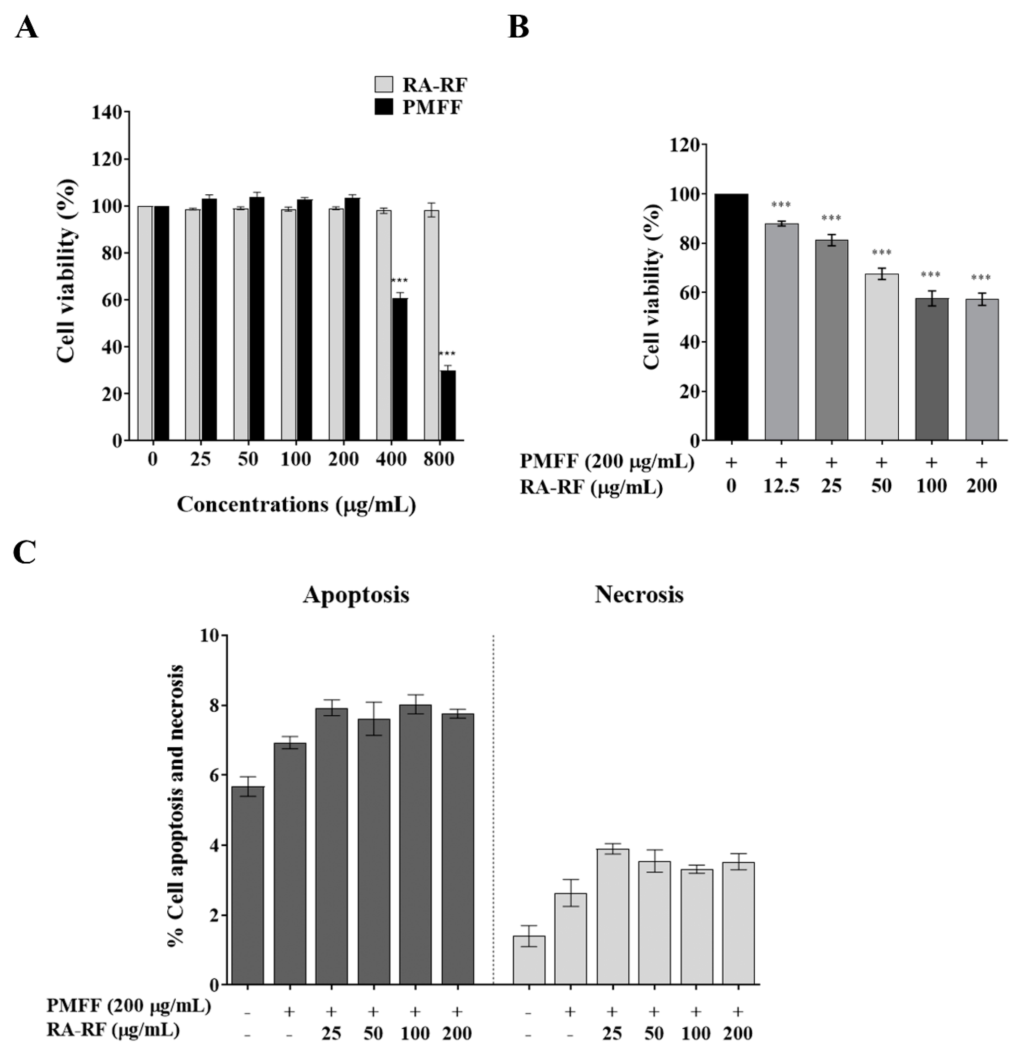
3.3. RA-RF Enhances Cell Viability and Apoptosis of PMFF-Induced A549 Cells

As shown in Figure 1A, the MTT assay revealed that cell viability was observed after treating RA-RF (0–800 μg/mL). The toxic dose of PMFF was observed to be significant at 400–800 μg/mL when compared to the control. Cell viability was not affected after exposure to RA-RF. As shown in Figure 1B, the A549 cells were pre-treated with RA-RF (0–200 µg/mL) and followed by a non-toxic dose of PMFF (200 µg/mL). Our results showed that the combination of RA-RF and PMFF significantly diminished cell viability in a dose-dependent manner. The 20% inhibitory concentration (IC20) of the co-treated mixture was approximately 25 µg/mL and the 50% inhibitory concentration (IC50) was higher than 200 µg/mL.
In addition, the apoptosis assay was used to confirm the cause of the cell viability reduction by the combination. As shown in Figure 1C, the combination of RA-RF and PMFF had no effect on cells apoptosis and necrosis compared to the control. The non-toxic concentrations of RA-RF in the range of 0–20 µg/mL were used for all of the following experiments.

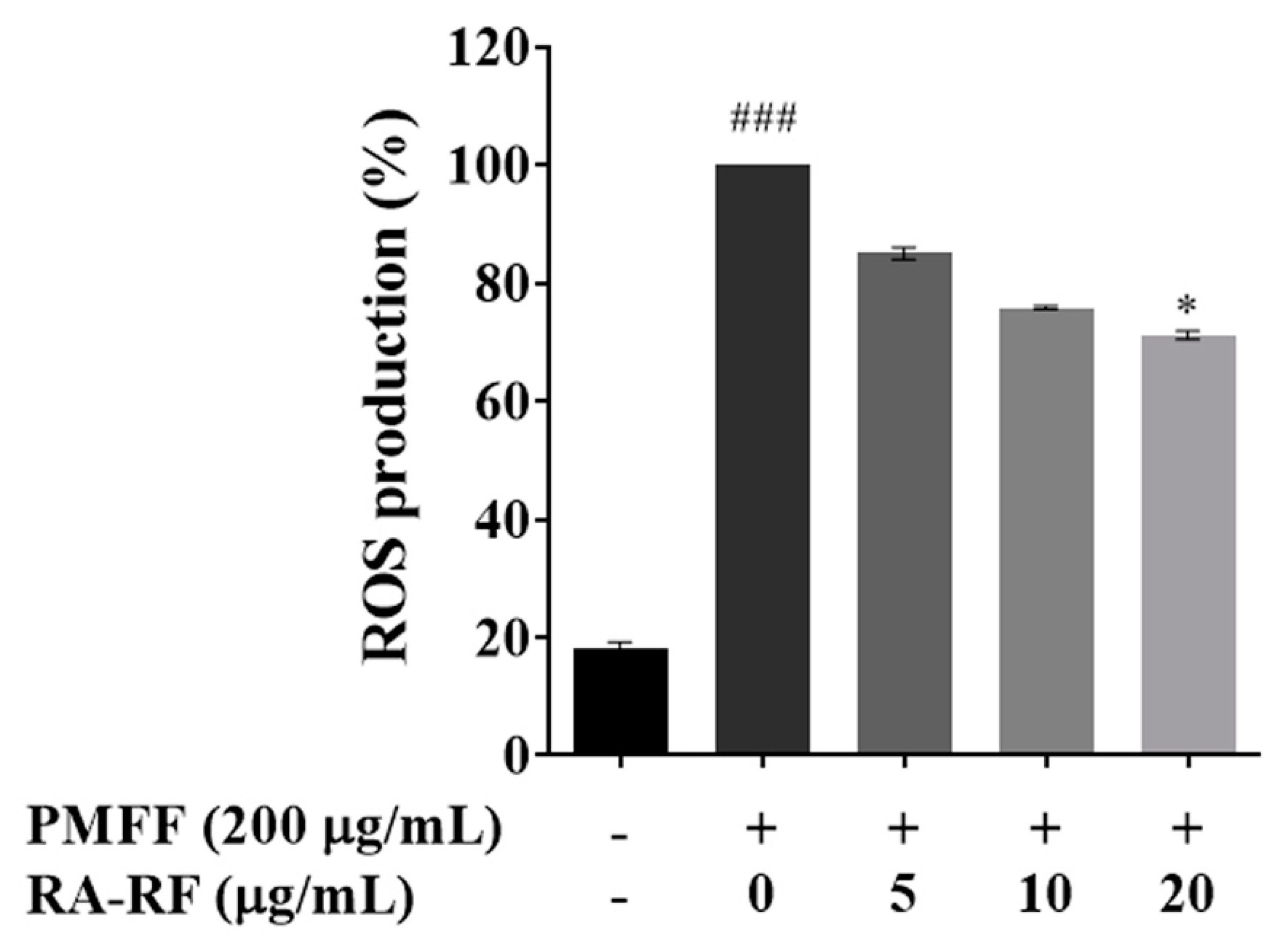
3.4. RA-RF Inhibits the Intracellular ROS Production

PM reportedly induces the production of ROS in A549 cells [35,36]. The effects of RA-RF on intracellular ROS production in PMFF-treated A549 cells were determined by DCFH-DA assay. As shown in Figure 2, our result demonstrated that PMFF drastically increased the level of ROS production approximately 5-fold (p < 0.001) compared to the control, whereas 20 µg/mL of RA-RF significantly decreased the percentage of ROS formation (p < 0.05). This result strongly suggested that RA-RF could inhibit intracellular ROS induction, which typically causes cellular and oxidative damage in PMFF-induced A549 cells.

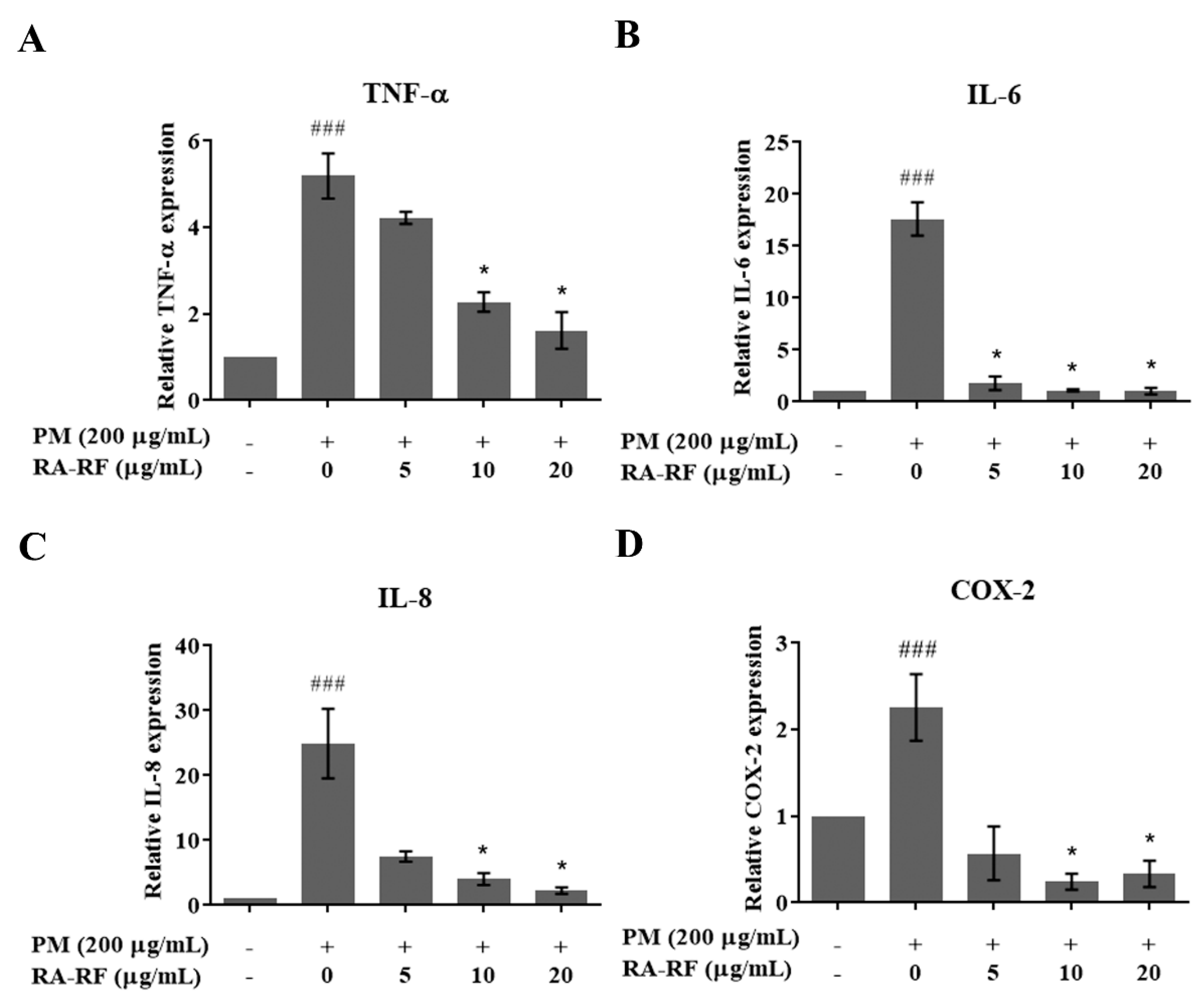
3.5. RA-RF Reducing the Inflammatory Cytokines and COX-2 Transcript

PM induces oxidative stress, triggering inflammatory cytokines such as IL-6, IL-8, TNF-α, and COX2 enzyme [11,37]. As shown in Figure 3A–D, PMFF considerably induced the mRNA expression of TNF-α, IL-6, IL-8, and COX2, whereas RA-RF-treated cells showed significantly decreased expression of those transcripts. This result revealed that RA-RF could reduce the regulation of inflammatory cytokines and COX2 enzyme induced by PMFF.

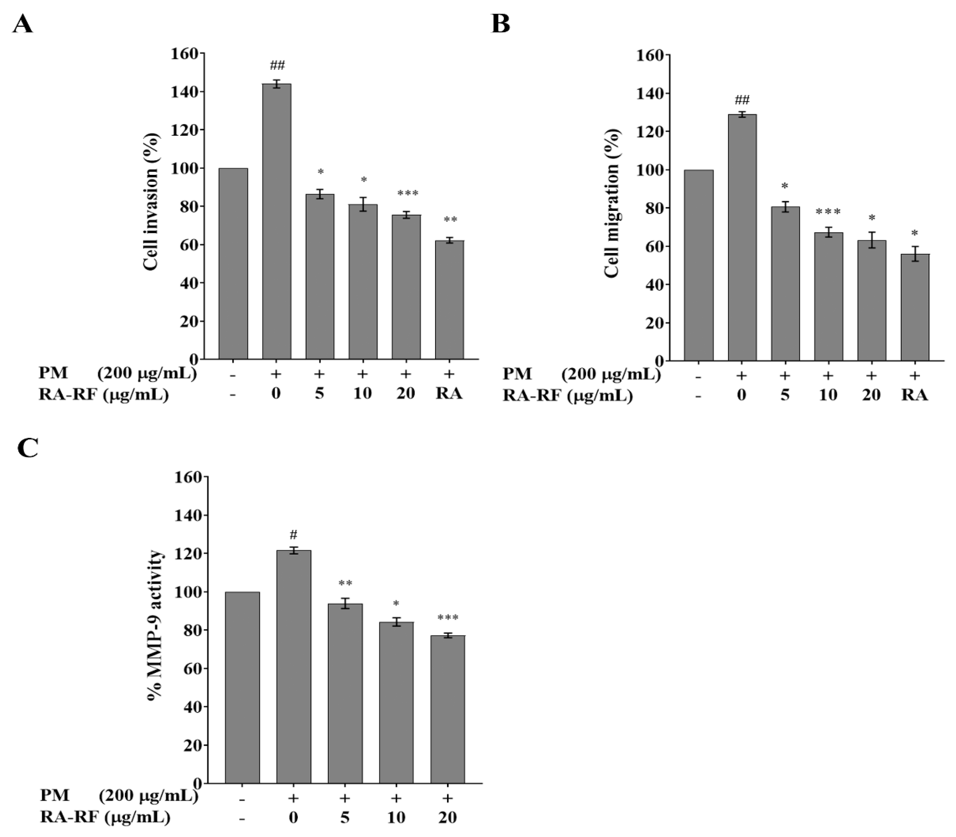
3.6. RA-RF Inhibits Invasion, Migration and MMP-9 Activity of A549 Cells Induced by PMFF

PM plays an important role in lung cancer formation and metastasis [13]. In this study, the anti-metastasis of RA-RF on PM-treated cells was investigated using Matrigel invasion and Transwell migration assays. As shown in Figure 4A, the invasive cells with PMFF treatment were increased by 1.44-fold (p < 0.01) compared to the non-treated cells. This revealed that treatment with 5, 10, and 20 μg/mL RA-RF significantly inhibited the invasion of PMFF-treated cells in a dose-dependent manner compared with commercial RA (5 μg/mL). Meanwhile, PMFF-treated cell migration significantly increased to 1.29-fold (p < 0.01); conversely, RA-RF (5–20 µg/mL) treatment significantly decreased PM-mediated migration in a concentration-dependent pattern compared to 5 μg/mL commercial RA (Figure 4B). Moreover, RA-RF (5–20 µg/mL) considerably reduced MMP-9 activity via a dose–response relationship (Figure 4C). In summary, polyphenols in RA-RF are capable of suppressing the invasion, migration, and MMP-9 activity of PMFF-induced A549 cells.

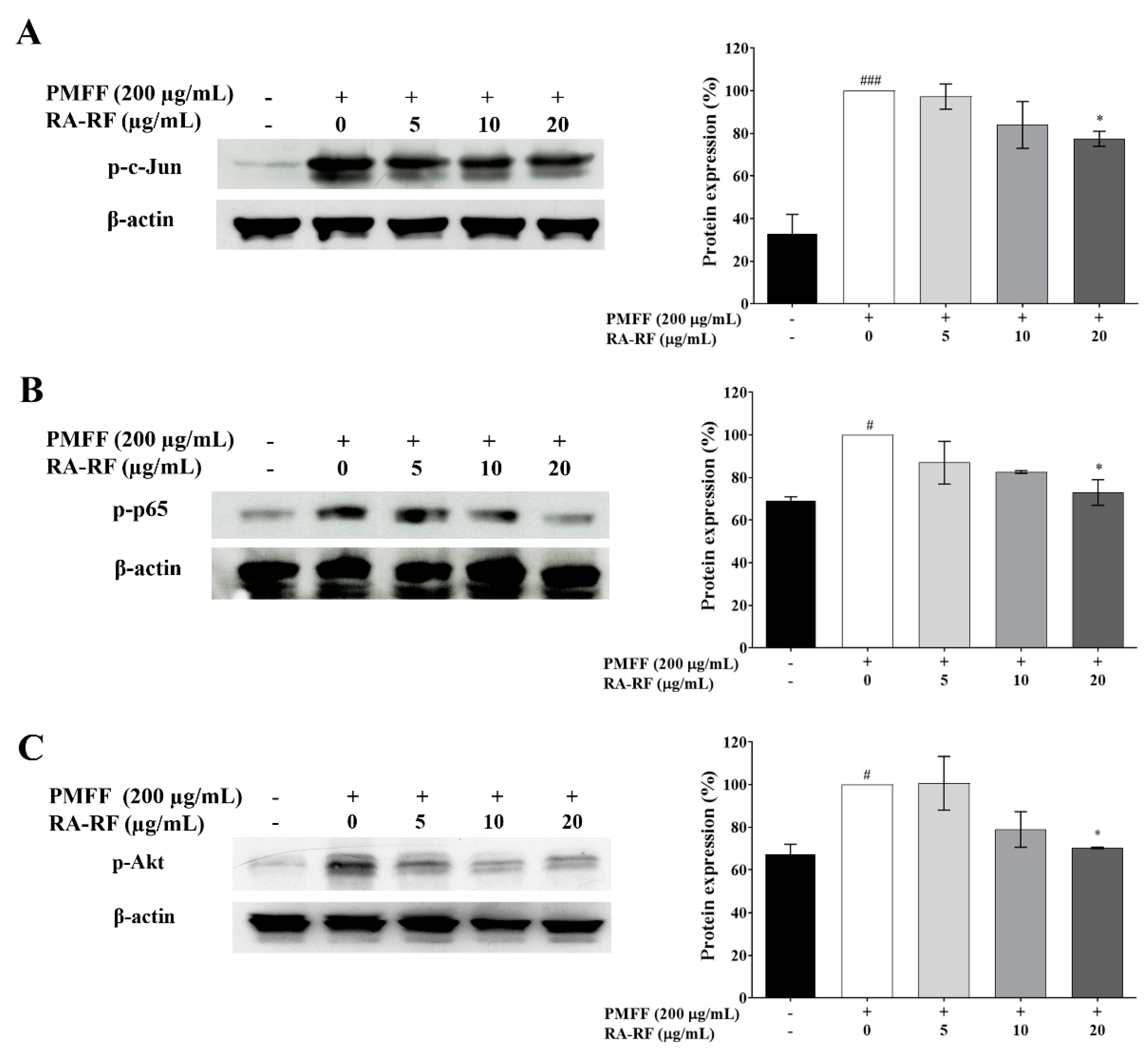
3.7. RA-RF Diminishes AP1, NF-κB and Akt Signaling Pathways

It has been reported the ROS-induced inflammation and metastasis are mediated through AP-1, NF-κB, and Akt signaling [38]. Therefore, this study determined the inducing effect of PMFF and the inhibitory effect of RA-RF on AP-1 (c-Jun), NF-κB (p-65), and Akt phosphorylation in A549 cells via Western blot analysis. As shown in Figure 5A–C, PMFF induced the phosphorylation of c-Jun, p-65-NF-κB, and Akt, whereas RA-RF (20 µg/mL) significantly inhibited the expression level of these signaling molecules in PMFF-treated cells. Collectively, RA-RF potentially inhibited the c-Jun, p-65-NF-κB, and Akt signaling pathways.

4. Discussion

Epidemiological studies have recently shown that PM is a crucial environmental contaminant related to various diseases such as pulmonary disease, cardiovascular disease, and lung cancer due to its ability to activate various inflammatory signaling pathways [39]. PM can also trigger both in vitro and in vivo determinations of the inflammatory response [3,40], metastasis induction [41], and other respiratory diseases [4]. Among several chemical components such as PAHs, metals, and trace elements, it was found that PAHs are predominantly absorbed in PM [39,42,43]. The PMFF samples were analyzed, and the concentrations of the 15 measured PAHs ranged from 0 to 2.094 ng/m3, with the highest component of PAHs being 2.094 ng/m3 of benzo[ghi]perylene, followed by, in order, indeno[1,2,3,-cd]pyrene, benzo[b]fluoranthene, and benzo[a]pyrene. Similarly, several studies in northern Thailand found that PM-bound PAHs were highly contaminated in the burning period [26,44,45,46,47].
These major PAH compounds were consistent with the data in our study. However, the PAH concentrations were lower than the values reported by Pooltawee et al. [45] in March 2014 in Phayao (nearly 180 ng/m3) and higher than those of the other studies in Chiangmai and Lamphun (0–1.98 ng/m3) [46,47]. This difference might be due to the period times, the area of PM collection, climate conditions such as season, temperature, rain, and humidity, and the scale of open burning derived from a number of hotspots [46,47].
To determine whether exposure to PAHs might cause significant effects in humans, health risk assessments can be calculated from the toxicity equivalent concentration (TEQ) based on PAH concentrations and toxic equivalent factors (TEFs) [26,48,49]. We found that the TEQ values in our study by using two equations as references from U.S. EPA (1993) [48] and Cecinato (1997) [49] were 1.37 ng/m3 and 1.69 ng/m3, respectively. These results were considerably higher than values reported by Wiriya W et al. and Yang N et al. [46,47]. In addition, the inhalation cancer risk (ICR) assessment was used for estimating the value of cancer risk from PAHs exposure by the equation ICR = TEQ × IURBaP [26,50] and IURBaP is the inhalation unit of risk defined as the risk of cancer from a lifetime (70 years) of inhalation of the unit mass of BaP, which is recommended as 8.7 × 10⁻² m3/μg by the World Health Organization (WHO, 2000) [51]. The ICR values > 10−4 are potential cancer risk [26,52] and the values of our study were in the range of 1.19 × 10⁻⁴ and 1.47 × 10⁻⁴, which are similar to the previous report in Nan province, Thailand, in 2018 (1.01 × 10⁻⁴ and 1.33 × 10⁻⁴) [26], indicating potential cancer risk.
Based on the International Agency for Research on Cancer (IARC), PAHs are classified as carcinogenic, probably carcinogenic (class 1, 2A or 2B), and not classifiable (class 3) to humans [53]. Interestingly, our results detected all PAHs of high molecular weight, including benzo[a]pyrene (class 1), dibenz[a,h]anthracene (class 2A), benz[a]anthracene, benzo[b]fluoranthene, benzo[k]fluoranthene, chrysene, Indeno[1,2,3-cd]pyrene (class 2B), benzo[ghi]perylene, fluoranthene, and pyrene (class 3). The health effects of human inhalation exposure to PMFF containing PAHs from fire haze could induce oxidative stress, inflammation, invasion, migration, and risk of lung cancer [13,54,55,56]. Furthermore, these harmful effects might be also caused by metals, endotoxins, and other elements in PMFF [57,58]
In this investigation, PAH content in PMFF, cytotoxicity, ROS, pro-inflammatory cytokines, invasion and migration effects, and the molecular mechanism of RA-RF on A549 cells induced by PMFF were determined. In the cell viability test, we found that RA-RF (0–800 μg/mL) and PMFF (0–200 μg/mL) had no effect on cytotoxicity, but the high concentrations of PMFF (400 and 800 μg/mL) significantly decreased cell survival. The cytotoxic effects of PMFF may depend on the concentration of PAHs, metals, endotoxins, and other elements [57,58]. Interestingly, co-treated PMFF with RA-RF had no effect on cell apoptosis and necrosis. The cell death types of A549 exposed to PMFF may be autophagy and should be further investigated.
Oxidative stress is a consequence of the excessive ROS activity generated while antioxidant defenses are suppressed [59]. Numerous reports have demonstrated that oxidative stress plays an important role in the cytotoxicity, inflammatory response, and carcinogenesis of the PM-induced cells [58,60,61,62]. The studies have shown that PM exposure increases intracellular ROS generation in human umbilical vein vascular endothelial cells [63], human microvascular endothelial cells [64], and human adenocarcinoma A549 cells [65].
In the current study, we investigated the effect of RA-RF polyphenol on alleviating the production of ROS treated by PMFF in A549 cells. Our result illustrated that PMFF induced the level of ROS production, which is consistent with previous studies showing a significant increase in the ROS level in A549 cells after exposure to PM [66,67]. Importantly, non-cytotoxic doses of RA-RF significantly suppressed the oxidative stress in PMFF-induced A549 cells by diminishing ROS production. These findings are correlated with our previous reports that the extracts from perilla reduced oxidative stress [22,68]. Additionally, RA decreased the level of ROS in RAW macrophages and asthma mouse models [69,70]. Collectively, our results indicate that RA-RF can suppress intracellular ROS generation in PMFF-stimulated A549 cells, and thus may protect against oxidative stress and injury in lung epithelial cells induced by air pollution.
It has been reported that high PM content is directly associated with an upregulation in ROS and proinflammatory responses [10,11,12]. Our experiment showed that PMFF increased the mRNA expression of IL-6, IL-8, TNF-α, and COX-2 in the treated cells. Consistent with the results of previous studies, PM from wildfire and wood smoke emissions produced high levels of IL-6, IL-8, TNF-α, and COX-2 both in vitro and in vivo [71,72,73]. Our results also demonstrated that RA-RF, a natural polyphenol component found in perilla, reduced IL-6, IL-8, TNF-α, and COX-2 gene expression. This finding is in agreement with our previous report showing that RA extracted from perilla reduced mRNA levels of pro-inflammatory cytokines [20,22,74]. Additionally, RA-rich extract from Trichodesma khasianum leaves could diminish ROS production and inflammatory modulators such as IL-6, TNF-α, and COX-2 levels in vitro and in vivo [75]. Commercial RA also alleviated IL-6, IL-8, TNF-α, and COX-2 levels in both cell and animal models [76,77,78,79,80].
In addition to the stimulation of ROS production, ROS overproduction is a distinctive feature of cancer progression and resistance to medical therapy. PM plays a crucial role in ROS hyperactivation and cancer metastasis and previous studies have shown that A549 cell invasion and migration were enhanced by PM [13,81]. Similarly, our results demonstrated that PMFF significantly induced the invasion, migration, and MMP-9 activity of A549 cells. On the other hand, these metastatic processes were suppressed dose-dependently by RA-RF. Our previous study found that the perilla leaves extract, which mainly contains RA and other polyphenols, strongly inhibits invasion and migration via diminished MMP-9 secretion and activity in breast cancer cells [19]. This result is consistent with other studies showing that the RA suppressed cancer cell invasion, migration, and MMP activity/expression for colon carcinoma cells [82], pancreatic cancer cells [83], liver cancer cells [84], and human glioma cells [85].
Several studies have demonstrated PM-induced pulmonary inflammation via oxidative stress, autophagy, and cell apoptosis [86]. Moreover, PM-induced overproduction of ROS plays an important role in the inflammatory process and promotes lung cancer metastasis [6]. Therefore, controlling the oxidative stress may be a potential target for preventing or reducing PM-induced pulmonary inflammation and lung cancer metastasis.
The AP-1 is a ubiquitous dimeric protein complex of c-Jun and Fos. The expression level and phosphorylation in post-translation modifications regulate the activity of AP-1 [87]. The inhibition of AP-1 activity has been shown to reduce inflammation [88], invasion, and migration [30]. Several studies have demonstrated that the phosphorylation of c-Jun can be induced by PM in many cell types such as human dermal fibroblasts [41], human keratinocytes cells [89], RAW macrophage cells [90], hepatocellular carcinoma cells [91], and human bronchial epithelial cells [92].
Our results found that PMFF enhanced c-Jun phosphorylation, whereas RA-RF suppressed the enhancement in A549 cells treated with PMFF. This work resembles previous studies showing that the ethanolic extract of perilla leaves disrupted c-Jun phosphorylation [93]. Moreover, RA-rich extract from perilla seed meal could reduce RANKL-induced c-Jun translocation in RAW macrophage cells [74]. In addition, RA alone exerted protective effects against oxidative stress, inflammation, invasion, migration, and MMP-9 protein expression through the AP-1 signaling cascade [94,95].
NF-kB is an inducible transcription factor regulating a set of genes involved in inflammatory responses and cancer cell migration [96]. The classic NF-kB is composed of RelA (p65)/c-Rel heterodimers. This complex was phosphorylated and translocated to the nucleus for activating the expression of the target genes [97]. Recent studies show that the NF-κB signaling cascade can be activated via high production of ROS stimulating the inflammation and immune response, and this activation can also develop the migration ability of human lung carcinoma [98,99]. Additionally, NF-κB is also responsible for the inflammatory and metastatic effects of PM in both in vitro and in vivo [11,100,101,102]. Here, we found that PMFF drastically induced the phosphorylation of p65-NF-κB in A549 cells. This finding is consistent with several previous studies showing that PM increased the phosphorylation of p65-NF-κB [90]. In our study, RA-RF evidently suppressed the phosphorylation of p65-NF-κB in A549 cells treated with PMFF. Other reports showed that the perilla extract rich in RA [74] and RA compounds had a considerable inhibitory effect on inflammation via suppression of the NF-κB signaling pathway during both in vitro and in vivo experiments [103,104]. Moreover, RA inhibited the invasion and migration of human glioma cells [85] and human hepatoma cells through the PI3K/Akt/NF-κB signaling pathway [105].
Furthermore, it is well known that the PI3K/Akt signaling pathway plays an important role in cell inflammation, cell apoptosis, angiogenesis, and metastasis [106]. PM activated ROS production and the Akt signaling pathway, leading to inflammation of the human bronchial epithelial B2B cell line [107], invasion, and migration of hepatocellular carcinoma cells [108]. We have shown that PMFF augmented Akt phosphorylation but RA-RF suppressed the phosphorylation activity in PMFF-treated cells. Likewise, RA reduced the Akt signaling cascade, lowering TNF-α and IL-6 levels in rat models [79]. Moreover, RA suppressed the PDPK1/Akt/mTOR pathways via LPS-induced neuroinflammation both in vitro and in vivo [109]. Moreover, the invasion and migration of cancer cells were inhibited by RA through the Akt signaling cascade [110,111,112].

5. Conclusions

The inhibitory effects of RA-RF on oxidative stress, inflammation, and metastasis caused by PMFF induction in cancer cells have never been reported. Our report is the first to show that RA-RF from perilla seed meal exerted anti-oxidative stress, anti-inflammatory, and anti-metastasis effects on PMFF-induced A549 cells. It is concluded that RA-RF markedly suppressed the ROS production induced by PMFF, resulting in reduced IL-6, IL-8, TNF-α, and COX-2 mRNA expression and inhibited the metastasis cascade affecting MMP-9 activity, which reduced cancer cell invasion and migration via the AP1, NF-κB, and Akt signaling pathways. However, further in vivo study of RA-RF should be performed to explore its use as an inhalation agent for preventing lung inflammation and cancer metastasis induced by PM exposure.

Author Contributions

Conceptualization, K.P., M.S., P.T. and W.C.; methodology, K.P., P.T. and W.C.; validation, K.P., P.T., S.Y. and W.C.; formal analysis, K.P., P.T., S.Y. and W.C.; investigation, K.P., P.T. and W.C.; resources, K.P. and P.T.; data curation, K.P., P.T. and W.C.; writing—original draft preparation, K.P., P.T.; writing—review and editing, K.P., P.T., S.Y. and W.C.; visualization, K.P. and P.T.; supervision, M.S.; project administration, K.P., P.T. and W.C.; funding acquisition, K.P., P.T. All authors have read and agreed to the published version of the manuscript.

Funding

This research was funded by the Thailand Research Fund (TRF) and Office of the Higher Education Commission (OHEC) (grant number MRG6180080), University of Phayao (Unit of Excellence grant number FF64-UoE019), and University of Phayao research grant number RD62055.

Institutional Review Board Statement

Not applicable.

Informed Consent Statement

Not applicable.

Data Availability Statement

Not applicable.

Acknowledgments

The authors wish to acknowledge the School of Medical Sciences, University of Phayao and the Faculty of Medicine, Chiang Mai University and Plant Genetic Conservation Project under the Royal Initiative of Her Royal Highness Princess Maha Chakri Sirindhorn for the use of their facilitates and support.

Conflicts of Interest

The authors declare no conflict of interest.

References

  1. Mueller, W.; Loh, M.; Vardoulakis, S.; Johnston, H.J.; Steinle, S.; Precha, N.; Kliengchuay, W.; Tantrakarnapa, K.; Cherrie, J.W. Ambient particulate matter and biomass burning: An ecological time series study of respiratory and cardiovascular hospital visits in northern Thailand. Environ. Health 2020, 19, 77. [Google Scholar] [CrossRef] [PubMed]
  2. Phairuang, W.; Hata, M.; Furuuchi, M. Influence of agricultural activities, forest fires and agro-industries on air quality in Thailand. J. Environ. Sci. 2017, 52, 85–97. [Google Scholar] [CrossRef] [PubMed]
  3. Farina, F.; Sancini, G.; Battaglia, C.; Tinaglia, V.; Mantecca, P.; Camatini, M.; Palestini, P. Milano summer particulate matter (PM10) triggers lung inflammation and extra pulmonary adverse events in mice. PLoS ONE 2013, 8, e56636. [Google Scholar] [CrossRef] [PubMed] [Green Version]
  4. Kim, K.; Kabir, E.; Kabir, S. A review on the human health impact of airborne particulate matter. Environ. Int. 2015, 74, 136–143. [Google Scholar] [CrossRef] [PubMed]
  5. Jakovljević, I.; Pehnec, G.; Vađić, V.; Čačković, M.; Tomašić, V.; Jelinić, J.D. Polycyclic aromatic hydrocarbons in PM10, PM2.5 and PM1 particle fractions in an urban area. Air Qual. Atmos. Health 2018, 11, 843–854. [Google Scholar] [CrossRef]
  6. Valavanidis, A.; Vlachogianni, T.; Fiotakis, K.; Loridas, S. Pulmonary oxidative stress, inflammation and cancer: Respirable particulate matter, fibrous dusts and ozone as major causes of lung carcinogenesis through reactive oxygen species mechanisms. J. Environ. Res. Public Health 2013, 10, 3886. [Google Scholar] [CrossRef] [PubMed]
  7. Freitas, M.; Alves, V.; Sarmento-Ribeiro, A.; Mota-Pinto, A. Polycyclic aromatic hydrocarbons may contibute for prostate cancer progression. J. Cancer Ther. 2013, 4, 37–46. [Google Scholar] [CrossRef] [Green Version]
  8. Yang, H.; Yang, T.; Gowrisankar, Y.V.; Liao, C.; Liao, J.; Huang, P.; Hseu, Y. Suppression of LPS-induced inflammation by chalcone flavokawain a through activation of Nrf2/ARE-mediated antioxidant genes and inhibition of ROS/NFκB signaling pathways in primary splenocytes. Oxid. Med. Cell. Longev. 2020, 3476212. [Google Scholar] [CrossRef]
  9. Kim, A.; Im, M.; Yim, N.; Jung, Y.P.; Ma, J.Y. Aqueous extract of bambusae caulis in taeniam inhibits PMA-induced tumor cell invasion and pulmonary metastasis: Suppression of NF-κB activation through ROS Signaling. PLoS ONE 2013, 8, e78061. [Google Scholar] [CrossRef]
  10. Sanjeewa, K.K.A.; Jayawardena, T.U.; Kim, S.-Y.; Lee, H.G.; Je, J.-G.; Jee, Y.; Jeon, Y.-J. Sargassum horneri (Turner) inhibit urban particulate matter-induced inflammation in MH-S lung macrophages via blocking TLRs mediated NF-κB and MAPK activation. J. Ethnopharmacol. 2020, 249, 112363. [Google Scholar] [CrossRef]
  11. Wang, J.; Huang, J.; Wang, L.; Chen, C.; Yang, D.; Jin, M.; Bai, C.; Song, Y. Urban particulate matter triggers lung inflammation via the ROS-MAPK-NF-κB signaling pathway. J. Thorac. Dis. 2017, 9, 4398–4412. [Google Scholar] [CrossRef] [PubMed] [Green Version]
  12. Cheng, W.; Lu, J.; Wang, B.; Sun, L.; Zhu, B.; Zhou, F.; Ding, Z. Inhibition of inflammation-induced injury and cell migration by coelonin and militarine in PM2.5-exposed human lung alveolar epithelial A549 cells. Eur. J. Pharmacol. 2021, 896, 173931. [Google Scholar] [CrossRef]
  13. Yue, H.; Yun, Y.; Gao, R.; Li, G.; Sang, N. Winter polycyclic aromatic hydrocarbon-bound particulate matter from peri-urban north china promotes lung cancer cell metastasis. Environ. Sci. Technol. 2015, 49, 14484–14493. [Google Scholar] [CrossRef]
  14. Vo, T.T.T.; Lee, C.; Wu, C.; Liu, J.; Lin, W.; Chen, Y.; Hsu, L.; Tsai11, M.; Lee, I. Surfactin from Bacillus subtilis attenuates ambient air particulate matter-promoted human oral cancer cells metastatic potential. J. Cancer 2020, 11, 6038–6049. [Google Scholar] [CrossRef]
  15. Seok, J.K.; Lee, J.; Kim, Y.M.; Boo, Y.C. Punicalagin and (–)-Epigallocatechin-3-Gallate rescue cell viability and attenuate inflammatory responses of human epidermal keratinocytes exposed to airborne particulate matter PM10. Skin Pharmacol. Physiol. 2018, 31, 134–143. [Google Scholar] [CrossRef] [PubMed]
  16. Boo, Y.C. Can plant phenolic compounds protect the skin from airborne particulate matter? Antioxidants 2019, 8, 379. [Google Scholar] [CrossRef] [Green Version]
  17. Ahmed, H.M. Ethnomedicinal, phytochemical and pharmacological investigations of Perilla frutescens (L.) Britt. Molecules 2019, 24, 102. [Google Scholar] [CrossRef] [PubMed] [Green Version]
  18. Suttajit, M.; Khanaree, C.; Tantipaiboonwong, P.; Pintha, K. Omega-3, omega-6 fatty acids and nutrients of Nga-mon seeds in northern Thailand. Naresuan Phayao J. 2015, 8, 80–86. [Google Scholar]
  19. Pintha, K.; Tantipaiboonwong, P.; Yodkeeree, S.; Chaiwangyen, W.; Chumphukam, O.; Khantamat, O.; Khanaree, C.; Kangwan, N.; Thongchuai, B.; Suttajit, M.; et al. Thai perilla (Perilla frutescens) leaf extract inhibits human breast cancer invasion and migration. Maejo Int. J. Sci. 2018, 12, 112–123. [Google Scholar]
  20. Kangwan, N.; Pintha, K.; Lekawanvijit, S.; Suttajit, M. Rosmarinic acid enriched fraction from Perilla frutescens leaves strongly protects indomethacin-induced gastric ulcer in rats. BioMed Res. Int. 2019, 2019, 9514703. [Google Scholar] [CrossRef] [Green Version]
  21. Khanaree, C.; Pintha, K.; Tantipaiboonwong, P.; Suttajit, M.; Chewonarin, T. The effect of Perilla frutescens leaf on 1, 2-dimethylhydrazine-induced initiation of colon carcinogenesis in rats. J. Food Biochem. 2018, 42, e12493. [Google Scholar] [CrossRef]
  22. Chumphukam, O.; Pintha, K.; Khanaree, C.; Chewonarin, T.; Chaiwangyen, W.; Tantipaiboonwong, P.; Suttajit, M.; Khantamat, O. Potential anti-mutagenicity, antioxidant, and anti-inflammatory capacities of the extract from perilla seed meal. J. Food Biochem. 2018, 42, e12556. [Google Scholar] [CrossRef]
  23. Shekarchi, M.; Hajimehdipoor, H.; Saeidnia, S.; Gohari, A.R.; Hamedani, M.P. Comparative study of rosmarinic acid content in some plants of Labiatae family. Pharmacogn. Mag. 2012, 8, 37–41. [Google Scholar]
  24. Kaewnarin, K.; Shank, L.; Niamsup, H.; Rakariyatham, N. Inhibitory effects of Lamiaceae plants on the formation of advanced glycation endproducts (AGEs) in model proteins. J. Med. Biol. Eng. 2013, 2, 224–227. [Google Scholar] [CrossRef]
  25. Sik, B.; Hanczné, E.L.; Kapcsándi, V.; Ajtony, Z. Conventional and nonconventional extraction techniques for optimal extraction processes of rosmarinic acid from six Lamiaceae plants as determined by HPLC-DAD measurement. J. Pharm. Biomed. Anal. 2020, 184, 113173. [Google Scholar] [CrossRef]
  26. Yabueng, N.; Wiriya, W.; Chantara, S. Influence of zero-burning policy and climate phenomena on ambient PM2.5 patterns and PAHs inhalation cancer risk during episodes of smoke haze in Northern Thailand. Atmos. Environ. 2020, 232, 117485. [Google Scholar] [CrossRef]
  27. Kim, E.J.; Kim, G.T.; Kim, B.M.; Lim, E.G.; Kim, S.-Y.; Kim, Y.M. Apoptosis-induced effects of extract from Artemisia annua Linné by modulating PTEN/p53/PDK1/Akt/ signal pathways through PTEN/p53-independent manner in HCT116 colon cancer cells. BMC Complement. Altern. Med. 2017, 17, 236. [Google Scholar] [CrossRef] [PubMed] [Green Version]
  28. Livak, K.J.; Schmittgen, T.D. Analysis of relative gene expression data using real-time quantitative PCR and the 2−ΔΔCT method. Methods 2001, 25, 402–408. [Google Scholar] [CrossRef]
  29. Chaiwangyen, W.; Ospina-Prieto, S.; Photini, S.M.; Schleussner, E.; Markert, U.R.; Morales-Prieto, D.M. Dissimilar microRNA-21 functions and targets in trophoblastic cell lines of different origin. Int. J. Biochem. Cell. Biol. 2015, 68, 187–196. [Google Scholar] [CrossRef] [PubMed]
  30. Ooppachai, C.; Limtrakul, P.; Yodkeeree, S. Dicentrine potentiates TNF-α-induced apoptosis and suppresses invasion of A549 lung adenocarcinoma cells via modulation of NF-κB and AP-1 activation. Molecules 2019, 24, 4100. [Google Scholar] [CrossRef] [Green Version]
  31. Kasala, E.R.; Bodduluru, L.N.; Barua, C.C.; Sriram, C.S.; Gogoi, R. Benzo(a)pyrene induced lung cancer: Role of dietary phytochemicals in chemoprevention. Pharmacol. Rep. 2015, 67, 996–1009. [Google Scholar] [CrossRef] [PubMed]
  32. Kim, K.H.; Jahan, S.A.; Kabir, E.; Brown, R.J. A review of airborne polycyclic aromatic hydrocarbons (PAHs) and their human health effects. Environ. Int. 2013, 60, 71–80. [Google Scholar] [CrossRef] [PubMed]
  33. Zhang, C.; Luo, Y.; Zhong, R.; Law, P.T.Y.; Boon, S.S.; Chen, Z.; Wong, C.H.; Chan, P.K.S. Role of polycyclic aromatic hydrocarbons as a co-factor in human papillomavirus-mediated carcinogenesis. BMC Cancer 2019, 19, 138. [Google Scholar] [CrossRef]
  34. Alagawany, M.; Abd El-Hack, M.E.; Farag, M.R.; Gopi, M.; Karthik, K.; Malik, Y.S.; Dhama, K. Rosmarinic acid: Modes of action, medicinal values and health benefits. Anim. Health Res. Rev. 2017, 18, 167. [Google Scholar] [CrossRef]
  35. Yi, S.; Zhang, F.; Qu, F.; Ding, W. Water-insoluble fraction of airborne particulate matter (PM10) induces oxidative stress in human lung epithelial A549 cells. Environ. Toxicol. 2014, 29, 226–233. [Google Scholar] [CrossRef]
  36. Yun, Y.; Gao, R.; Yue, H.; Li, G.; Zhu, N.; Sang, N. Synergistic effects of particulate matter (PM10) and SO2 on human non-small cell lung cancer A549 via ROS-mediated NF-κB activation. J. Environ. Sci. 2015, 31, 146–153. [Google Scholar] [CrossRef]
  37. Kim, K.E.; Cho, D.; Park, H.J. Air pollution and skin diseases: Adverse effects of airborne particulate matter on various skin diseases. Life Sci. 2016, 152, 126–134. [Google Scholar] [CrossRef]
  38. Lee, G.H.; Jin, S.W.; Kim, S.J.; Pham, T.H.; Choi, J.H.; Jeong, H.G. Tetrabromobisphenol A induces MMP-9 expression via NADPH oxidase and the activation of ROS, MAPK, and Akt pathways in human breast cancer MCF-7 Cells. Toxicol. Res. 2019, 35, 93–101. [Google Scholar] [CrossRef]
  39. Yan, Z.; Jin, Y.; An, Z.; Liu, Y.; Samet, J.M.; Wu, W. Inflammatory cell signaling following exposures to particulate matter and ozone. Biochim. Biophys. Acta 2016, 1860, 2826–2834. [Google Scholar] [CrossRef] [PubMed]
  40. Radan, M.; Dianat, M.; Badavi, M.; Mard, S.A.; Bayati, V.; Goudarzi, G. In vivo and in vitro evidence for the involvement of Nrf2-antioxidant response element signaling pathway in the inflammation and oxidative stress induced by particulate matter (PM10): The effective role of gallic acid. Free Radic. Res. 2019, 53, 210–225. [Google Scholar] [CrossRef]
  41. Li, W.; Liu, T.; Xiong, Y.; Lv, J.; Cui, X.; He, R. Diesel exhaust particle promotes tumor lung metastasis via the induction of BLT1-mediated neutrophilic lung inflammation. Cytokine 2018, 111, 530–540. [Google Scholar] [CrossRef] [PubMed]
  42. Jaafari, J.; Naddafi, K.; Yunesian, M.; Nabizadeh, R.; Hassanvand, M.S.; Ghozikali, M.G.; Shamsollahi, H.R.; Nazmara, S.; Yaghmaeian, K. Characterization, risk assessment and potential source identification of PM10 in Tehran. Microchem. J. 2020, 154, 104533. [Google Scholar] [CrossRef]
  43. Kord Mostafapour, F.; Jaafari, J.; Gharibi, H.; Sepand, M.R.; Hoseini, M.; Balarak, D.; Sillanpää, M.; Javid, A.B. Characterizing of fine particulate matter (PM1) on the platforms and outdoor areas of underground and surface subway stations. Hum. Ecol. Risk Assess. 2018, 24, 1016–1029. [Google Scholar] [CrossRef]
  44. Pongpiachan, S.; Hattayanone, M.; Cao, J. Effect of agricultural waste burning season on PM2.5-bound polycyclic aromatic hydrocarbon (PAH) levels in Northern Thailand. Atmos. Pollut. Res. 2017, 8, 1069–1080. [Google Scholar] [CrossRef]
  45. Pooltawee, J.; Pimpunchat, B.; Junyapoon, S. Size distribution, characterization and risk assessment of particle-bound polycyclic aromatic hydrocarbons during haze periods in Phayao Province, northern Thailand. Air Qual. Atmos. Health 2017, 10, 1097–1112. [Google Scholar] [CrossRef]
  46. Wiriya, W.; Prapamontol, T.; Chantara, S. PM10-bound polycyclic aromatic hydrocarbons in Chiang Mai (Thailand): Seasonal variations, source identification, health risk assessment and their relationship to air-mass movement. Atmos. Res. 2013, 124, 109–122. [Google Scholar] [CrossRef]
  47. Pengchai, P.; Chantara, S.; Sopajaree, K.; Wangkarn, S.; Tengcharoenkul, U.; Rayanakorn, M. Seasonal variation, risk assessment and source estimation of PM 10 and PM10-bound PAHs in the ambient air of Chiang Mai and Lamphun, Thailand. Environ. Monit. Assess. 2008, 154, 197. [Google Scholar] [CrossRef]
  48. US Environmental Protection Agency. Provisional Guidance for Quantitative Risk Assessment of Polycyclic Aromatic Hydrocarbons; EPA-600/R-93/089; US Environmental Protection Agency: Research Triangle Park, NC, USA, 1993; Volume 600.
  49. Cecinato, A. Polynuclear aromatic hydrocarbons (PAH), benz (a) pyrene (BaPY) and nitrated-PAH (N-PAH) in suspended particulate matter: Proposal for revision of the Italian reference method. Ann. Chim. 1997, 87, 483–496. [Google Scholar]
  50. US Environmental Protection Agency. Guidelines for Carcinogen Risk Assessment. Risk Assessment Forum. Available online: https://www.epa.gov/risk/guidelines-carcinogen-risk-assessment (accessed on 20 July 2021).
  51. World Health Organization. Air Quality Guidelines for Europe; WHO Regional Office for Europe: Copenhagen, Denmark, 2000. [Google Scholar]
  52. Liao, C.; Chiang, K. Probabilistic risk assessment for personal exposure to carcinogenic polycyclic aromatic hydrocarbons in Taiwanese temples. Chemosphere 2006, 63, 1610–1619. [Google Scholar] [CrossRef] [PubMed]
  53. AIRC. Some non-heterocyclic polycyclic aromatic hydrocarbons and some related exposures. IARC Monogr. Eval. Carcinog. Risks Hum. 2010, 92, 1–853. [Google Scholar]
  54. Adetona, O.; Reinhardt, T.E.; Domitrovich, J.; Broyles, G.; Adetona, A.M.; Kleinman, M.T.; Ottmar, R.D.; Naeher, L.P. Review of the health effects of wildland fire smoke on wildland firefighters and the public. Inhal. Toxicol. 2016, 28, 95–139. [Google Scholar] [CrossRef]
  55. Eom, S.; Yim, D.; Moon, S.I.; Youn, J.; Kwon, H.; Oh, H.C.; Yang, J.J.; Park, S.K.; Yoo, K.; Kim, H.S.; et al. Polycyclic aromatic hydrocarbon-induced oxidative stress, antioxidant capacity, and the risk of lung cancer: A pilot nested case-control study. Anticancer Res. 2013, 33, 3089. [Google Scholar]
  56. Wickramasinghe, A.P.; Karunaratne, D.G.G.P.; Sivakanesan, R. PM10-bound polycyclic aromatic hydrocarbons: Biological indicators, lung cancer risk of realistic receptors and ‘source-exposure-effect relationship’ under different source scenarios. Chemosphere 2012, 87, 1381–1387. [Google Scholar] [CrossRef] [PubMed]
  57. Huang, H.; Tantoh, D.M.; Hsu, S.; Nfor, O.N.; Frank, C.L.; Lung, C.; Ho, C.; Chen, C.; Liaw, Y. Association between coarse particulate matter (PM10-2.5) and nasopharyngeal carcinoma among Taiwanese men. J. Investig. Med. 2020, 68, 419. [Google Scholar] [CrossRef] [PubMed] [Green Version]
  58. Danielsen, P.H.; Møller, P.; Jensen, K.A.; Sharma, A.K.; Wallin, H.; Bossi, R.; Autrup, H.; Mølhave, L.; Ravanat, J.-L.; Briedé, J.J.; et al. Oxidative stress, DNA damage, and inflammation induced by ambient air and wood smoke particulate matter in human A549 and THP-1 cell lines. Chem. Chem. Res. Toxicol. 2011, 24, 168–184. [Google Scholar] [CrossRef]
  59. Pisoschi, A.M.; Pop, A. The role of antioxidants in the chemistry of oxidative stress: A. review. Eur. J. Med. Chem. 2015, 97, 55–74. [Google Scholar] [CrossRef]
  60. Shang, Y.; Zhou, Q.; Wang, T.; Jiang, Y.; Zhong, Y.; Qian, G.; Zhu, T.; Qiu, X.; An, J. Airborne nitro-PAHs induce Nrf2/ARE defense system against oxidative stress and promote inflammatory process by activating PI3K/Akt pathway in A549 cells. Toxicol. Vitr. 2017, 44, 66–73. [Google Scholar] [CrossRef] [PubMed]
  61. Akhtar, U.S.; McWhinney, R.D.; Rastogi, N.; Abbatt, J.P.D.; Evans, G.J.; Scott, J.A. Cytotoxic and proinflammatory effects of ambient and source-related particulate matter (PM) in relation to the production of reactive oxygen species (ROS) and cytokine adsorption by particles. Inhal. Toxicol. 2010, 22, 37–47. [Google Scholar] [CrossRef] [PubMed]
  62. Das, A.; Habib, G.; Vivekanandan, P.; Kumar, A. Reactive oxygen species production and inflammatory effects of ambient PM2.5 -associated metals on human lung epithelial A549 cells “one year-long study”: The Delhi chapter. Chemosphere 2021, 262, 128305. [Google Scholar] [CrossRef]
  63. Long, Y.; Yang, X.; Yang, Q.; Clermont, A.C.; Yin, Y.; Liu, G.; Hu, L.; Liu, Q.; Zhou, Q.; Liu, Q.S.; et al. PM2.5 induces vascular permeability increase through activating MAPK/ERK signaling pathway and ROS generation. J. Hazard. Mater. 2020, 386, 121659. [Google Scholar] [CrossRef]
  64. Chua, M.L.; Setyawati, M.I.; Li, H.; Fang, C.H.Y.; Gurusamy, S.; Teoh, F.T.L.; Leong, D.T.; George, S. Particulate matter from indoor environments of classroom induced higher cytotoxicity and leakiness in human microvascular endothelial cells in comparison with those collected from corridor. Indoor Air 2017, 27, 551–563. [Google Scholar] [CrossRef]
  65. Lu, Y.; Su, S.; Jin, W.; Wang, B.; Li, N.; Shen, H.; Li, W.; Huang, Y.; Chen, H.; Zhang, Y.; et al. Characteristics and cellular effects of ambient particulate matter from Beijing. Environ. Pollut. 2014, 191, 63–69. [Google Scholar] [CrossRef]
  66. Danielsen, P.H.; Loft, S.; Kocbach, A.; Schwarze, P.E.; Møller, P. Oxidative damage to DNA and repair induced by Norwegian wood smoke particles in human A549 and THP-1 cell lines. Mutat. Res. 2009, 674, 116–122. [Google Scholar] [CrossRef]
  67. Libalova, H.; Milcova, A.; Cervena, T.; Vrbova, K.; Rossnerova, A.; Novakova, Z.; Topinka, J.; Rossner, P. Kinetics of ROS generation induced by polycyclic aromatic hydrocarbons and organic extracts from ambient air particulate matter in model human lung cell lines. Mutat Res. Genet. Toxicol. Environ. Mutagen. 2018, 827, 50–58. [Google Scholar] [CrossRef] [PubMed]
  68. Tipsuwan, W.; Chaiwangyen, W. Preventive effects of polyphenol-rich perilla leaves on oxidative stress and haemolysis. Sci. Asia 2018, 44, 162–169. [Google Scholar] [CrossRef] [Green Version]
  69. Qiao, S.; Li, W.; Tsubouchi, R.; Haneda, M.; Murakami, K.; Takeuchi, F.; Nisimoto, Y.; Yoshino, M. Rosmarinic acid inhibits the formation of reactive oxygen and nitrogen species in RAW264.7 macrophages. Free Radic. Res. 2005, 39, 995–1003. [Google Scholar] [CrossRef]
  70. Liang, Z.; Wu, L.; Deng, X.; Liang, Q.; Xu, Y.; Deng, R.; Lv, L.; Ji, M.; Hao, Z.; He, J. The antioxidant rosmarinic acid ameliorates oxidative lung damage in experimental allergic asthma via modulation of NADPH oxidases and antioxidant enzymes. Inflammation 2020, 43, 1902–1912. [Google Scholar] [CrossRef]
  71. Erlandsson, L.; Lindgren, R.; Nääv, Å.; Krais, A.M.; Strandberg, B.; Lundh, T.; Boman, C.; Isaxon, C.; Hansson, S.R.; Malmqvist, E. Exposure to wood smoke particles leads to inflammation, disrupted proliferation and damage to cellular structures in a human first trimester trophoblast cell line. Environ. Pollut. 2020, 264, 114790. [Google Scholar] [CrossRef]
  72. Young, T.M.; Black, G.P.; Wong, L.; Bloszies, C.S.; Fiehn, O.; He, G.; Denison, M.S.; Vogel, C.F.A.; Durbin-Johnson, B. Identifying toxicologically significant compounds in urban wildfire ash using in vitro bioassays and high-resolution mass spectrometry. Environ. Sci. Technol. 2021, 55, 3657–3667. [Google Scholar] [CrossRef]
  73. Williams, K.M.; Franzi, L.M.; Last, J.A. Cell-specific oxidative stress and cytotoxicity after wildfire coarse particulate matter instillation into mouse lung. Toxicol. Appl. Pharmacol. 2013, 266, 48–55. [Google Scholar] [CrossRef] [Green Version]
  74. Phromnoi, K.; Suttajit, M.; Saenjum, C.; Limtrakul, P.D. Inhibitory effect of a rosmarinic acid-enriched fraction prepared from Nga-Mon (Perilla frutescens) seed meal on osteoclastogenesis through the RANK signaling pathway. Antioxidants 2021, 10, 307. [Google Scholar] [CrossRef]
  75. Wang, G.; Chen, S.; Chen, Y.; Hong, C.; Hsu, Y.; Yen, G. Protective effect of rosmarinic acid-rich trichodesma khasianum clarke leaves against ethanol-induced gastric mucosal injury in vitro and in vivo. Phytomedicine 2021, 80, 153382. [Google Scholar] [CrossRef] [PubMed]
  76. Sadeghi, A.; Bastin, A.R.; Ghahremani, H.; Doustimotlagh, A.H. The effects of rosmarinic acid on oxidative stress parameters and inflammatory cytokines in lipopolysaccharide-induced peripheral blood mononuclear cells. Mol. Biol. Rep. 2020, 47, 3557–3566. [Google Scholar] [CrossRef] [PubMed]
  77. Lembo, S.; Balato, A.; Di Caprio, R.; Cirillo, T.; Giannini, V.; Gasparri, F.; Monfrecola, G. The modulatory effect of ellagic acid and rosmarinic acid on Ultraviolet-B-induced cytokine/chemokine gene expression in skin keratinocyte (HaCaT) cells. Biomed. Res. Int. 2014, 2014, 346793. [Google Scholar] [CrossRef] [PubMed]
  78. Wei, Y.; Chen, J.; Hu, Y.; Lu, W.; Zhang, X.; Wang, R.; Chu, K. Rosmarinic acid mitigates lipopolysaccharide-induced neuroinflammatory responses through the inhibition of TLR4 and CD14 expression and NF-κB and NLRP3 inflammasome activation. Inflammation 2018, 41, 732–740. [Google Scholar] [CrossRef]
  79. Rocha, J.; Eduardo-Figueira, M.; Barateiro, A.; Fernandes, A.; Brites, D.; Bronze, R.; Duarte, C.M.M.; Serra, A.T.; Pinto, R.; Freitas, M.; et al. Anti-inflammatory effect of rosmarinic acid and an extract of rosmarinus officinalis in rat models of local and systemic inflammation. Basic Clin. Pharmacol. Toxicol. 2015, 116, 398–413. [Google Scholar] [CrossRef]
  80. Domitrović, R.; Škoda, M.; Vasiljev Marchesi, V.; Cvijanović, O.; Pernjak Pugel, E.; Štefan, M.B. Rosmarinic acid ameliorates acute liver damage and fibrogenesis in carbon tetrachloride-intoxicated mice. Food Chem. Toxicol. 2013, 51, 370–378. [Google Scholar] [CrossRef]
  81. Morales-Bárcenas, R.; Chirino, Y.I.; Sánchez-Pérez, Y.; Osornio-Vargas, Á.R.; Melendez-Zajgla, J.; Rosas, I.; García-Cuellar, C.M. Particulate matter (PM10) induces metalloprotease activity and invasion in airway epithelial cells. Toxicol. Lett. 2015, 237, 167–173. [Google Scholar] [CrossRef]
  82. Han, Y.; Kee, J.; Hong, S. Rosmarinic acid activates ampk to inhibit metastasis of colorectal cancer. Front. Pharmacol. 2018, 9. [Google Scholar] [CrossRef]
  83. Han, Y.; Ma, L.; Zhao, L.; Feng, W.; Zheng, X. Rosmarinic inhibits cell proliferation, invasion and migration via up-regulating miR-506 and suppressing MMP2/16 expression in pancreatic cancer. Biomed. Pharmacother. 2019, 115, 108878. [Google Scholar] [CrossRef]
  84. Chen, X.; Su, H.Z.Z. Detailed studies on the anticancer action of rosmarinic acid in human Hep-G2 liver carcinoma cells: Evaluating its effects on cellular apoptosis, caspase activation and suppression of cell migration and invasion. J. BUON 2020, 25, 2011–2016. [Google Scholar] [PubMed]
  85. Liu, Y.; Xu, X.; Tang, H.; Pan, Y.; Hu, B.; Huang, G. Rosmarinic acid inhibits cell proliferation, migration, and invasion and induces apoptosis in human glioma cells. Int. J. Mol. Med. 2021, 47, 67. [Google Scholar] [CrossRef] [PubMed]
  86. Chan, Y.L.; Wang, B.; Chen, H.; Ho, K.F.; Cao, J.; Hai, G.; Jalaludin, B.; Herbert, C.; Thomas, P.S.; Saad, S.; et al. Pulmonary inflammation induced by low-dose particulate matter exposure in mice. Am. J. Physiol. Lung Cell. Mol. Physiol. 2019, 317, L424–L430. [Google Scholar] [CrossRef] [PubMed]
  87. Bejjani, F.; Evanno, E.; Zibara, K.; Piechaczyk, M.; Jariel-Encontre, I. The AP-1 transcriptional complex: Local switch or remote command? Biochim. Biophys. Acta Rev. Bioenergy Cancer 2019, 1872, 11–23. [Google Scholar] [CrossRef] [PubMed]
  88. Wang, L.; Lee, W.; Jayawardena, T.U.; Cha, S.; Jeon, Y. Dieckol, an algae-derived phenolic compound, suppresses airborne particulate matter-induced skin aging by inhibiting the expressions of pro-inflammatory cytokines and matrix metalloproteinases through regulating NF-κB, AP-1, and MAPKs signaling pathways. Food Chem. Toxicol. 2020, 146, 111823. [Google Scholar] [CrossRef]
  89. Kim, J.H.; Kim, M.; Kim, J.M.; Lee, M.K.; Seo, S.J.; Park, K.Y. Afzelin suppresses proinflammatory responses in particulate matter-exposed human keratinocytes. Int. J. Mol. Med. 2019, 43, 2516–2522. [Google Scholar] [CrossRef]
  90. Zu, Y.-Y.; Liu, Q.-F.; Tian, S.-X.; Jin, L.-X.; Jiang, F.-S.; Li, M.-Y.; Zhu, B.-Q.; Ding, Z.-S. Effective fraction of Bletilla striata reduces the inflammatory cytokine production induced by water and organic extracts of airborne fine particulate matter (PM2.5) in vitro. BMC Complement. Altern. Med. 2019, 19, 369. [Google Scholar] [CrossRef]
  91. Jarvis, I.W.H.; Bergvall, C.; Morales, D.A.; Kummrow, F.; Umbuzeiro, G.A.; Westerholm, R.; Stenius, U.; Dreij, K. Nanomolar levels of PAHs in extracts from urban air induce MAPK signaling in HepG2 cells. Toxicol. Lett. 2014, 229, 25–32. [Google Scholar] [CrossRef] [Green Version]
  92. Xu, F.; Luo, M.; He, L.; Cao, Y.; Li, W.; Ying, S.; Chen, Z.; Shen, H. Necroptosis contributes to urban particulate matter-induced airway epithelial injury. Cell. Physiol. Biochem. 2018, 46, 699–712. [Google Scholar] [CrossRef]
  93. Bae, J.; Han, M.; Shin, H.S.; Kim, M.; Shin, C.; Lee, D.H.; Chung, J.H. Perilla frutescens leaves extract ameliorates ultraviolet radiation-induced extracellular matrix damage in human dermal fibroblasts and hairless mice skin. J. Ethnopharmacol. 2017, 195, 334–342. [Google Scholar] [CrossRef]
  94. Lin, S.; Wang, Y.; Chen, W.; Liao, S.; Chou, S.; Yang, C.; Chen, C. Hepatoprotective activities of rosmarinic acid against extrahepatic cholestasis in rats. Food Chem. Toxicol. 2017, 108, 214–223. [Google Scholar] [CrossRef]
  95. Yang, K.; Shen, Z.; Zou, Y.; Gao, K. Rosmarinic acid inhibits migration, invasion, and p38/AP-1 signaling via miR-1225-5p in colorectal cancer cells. J. Recept. Signal. Transduct. Res. 2021, 41, 284–293. [Google Scholar] [CrossRef] [PubMed]
  96. Dolcet, X.; Llobet, D.; Pallares, J.; Matias-Guiu, X. NF-kB in development and progression of human cancer. Virchows Archiv 2005, 446, 475–482. [Google Scholar] [CrossRef] [PubMed]
  97. DiDonato, J.A.; Mercurio, F.; Karin, M. NF-κB and the link between inflammation and cancer. Immunol. Rev. 2012, 246, 379–400. [Google Scholar] [CrossRef] [PubMed]
  98. Gong, W.; Liu, J.; Yin, J.; Cui, J.; Xiao, D.; Zhuo, W.; Luo, C.; Liu, R.; Li, X.; Zhang, W.; et al. Resistin facilitates metastasis of lung adenocarcinoma through the TLR4/Src/EGFR/PI3K/NF-κB pathway. Cancer Sci. 2018, 109, 2391–2400. [Google Scholar] [CrossRef]
  99. Yahfoufi, N.; Alsadi, N.; Jambi, M.; Matar, C. The Immunomodulatory and Anti-Inflammatory Role of Polyphenols. Nutrients 2018, 10, 1618. [Google Scholar] [CrossRef] [Green Version]
  100. Cho, C.; Hsieh, W.; Tsai, C.; Chen, C.; Chang, H.; Lin, C. In vitro and in vivo experimental studies of PM2.5 on disease progression. Int. J. Environ. Res. Public Health 2018, 15, 1380. [Google Scholar] [CrossRef] [Green Version]
  101. Haghani, A.; Johnson, R.; Safi, N.; Zhang, H.; Thorwald, M.; Mousavi, A.; Woodward, N.C.; Shirmohammadi, F.; Coussa, V.; Wise, J.P.; et al. Toxicity of urban air pollution particulate matter in developing and adult mouse brain: Comparison of total and filter-eluted nanoparticles. Environ. Int. 2020, 136, 105510. [Google Scholar] [CrossRef]
  102. Taş, İ.; Zhou, R.; Park, S.-Y.; Yang, Y.; Gamage, C.D.B.; Son, Y.-J.; Paik, M.-J.; Kim, H. Inflammatory and tumorigenic effects of environmental pollutants found in particulate matter on lung epithelial cells. Toxicol. Vitr. 2019, 59, 300–311. [Google Scholar] [CrossRef]
  103. Joardar, S.; Dewanjee, S.; Bhowmick, S.; Dua, T.K.; Das, S.; Saha, A.; De Feo, V. Rosmarinic acid attenuates cadmium-induced nephrotoxicity via inhibition of oxidative stress, apoptosis, inflammation and fibrosis. Int. J. Mol. Sci. 2019, 20, 2027. [Google Scholar] [CrossRef] [Green Version]
  104. Cao, W.; Hu, C.; Wu, L.; Xu, L.; Jiang, W. Rosmarinic acid inhibits inflammation and angiogenesis of hepatocellular carcinoma by suppression of NF-κB signaling in H22 tumor-bearing mice. J. Pharmacol. Sci. 2016, 132, 131–137. [Google Scholar] [CrossRef] [Green Version]
  105. An, Y.; Zhao, J.; Zhang, Y.; Wu, W.; Hu, J.; Hao, H.; Qiao, Y.; Tao, Y.; An, L. Rosmarinic acid induces proliferation suppression of hepatoma cells associated with NF-κB signaling pathway.Asian Pac. J. Cancer Prev. 2020, 22, 1623–1632. [Google Scholar]
  106. Duan, M.; Zhou, H.; Wu, Q.; Liu, C.; Xiao, Y.; Deng, W.; Tang, Q. Andrographolide protects against hg-induced inflammation, apoptosis, migration, and impairment of angiogenesis via PI3K/AKT-eNOS Signalling in HUVECs. Mediators Inflamm. 2019, 2019, 6168340. [Google Scholar] [CrossRef]
  107. Song, L.; Li, D.; Li, X.; Ma, L.; Bai, X.; Wen, Z.; Zhang, X.; Chen, D.; Peng, L. Exposure to PM2.5 induces aberrant activation of NF-κB in human airway epithelial cells by downregulating miR-331 expression. Environ. Toxicol. Pharmacol. 2017, 50, 192–199. [Google Scholar] [CrossRef] [PubMed]
  108. Zhang, Q.; Luo, Q.; Yuan, X.; Chai, L.; Li, D.; Liu, J.; Lv, Z. Atmospheric particulate matter2.5 promotes the migration and invasion of hepatocellular carcinoma cells. Oncol. Lett. 2017, 13, 3445–3450. [Google Scholar] [CrossRef] [PubMed] [Green Version]
  109. Wei, Y.; Chen, J.; Cai, G.-E.; Lu, W.; Xu, W.; Wang, R.; Lin, Y.; Yang, C. Rosmarinic acid regulates microglial M1/M2 polarization via the PDPK1/Akt/HIF pathway under conditions of neuroinflammation. Inflammation 2021, 44, 129–147. [Google Scholar] [CrossRef]
  110. Wang, L.; Yang, H.; Wang, C.; Shi, X.; Li, K. Rosmarinic acid inhibits proliferation and invasion of hepatocellular carcinoma cells SMMC 7721 via PI3K/AKT/mTOR signal pathway. Biomed. Pharmacother. 2019, 120, 109443. [Google Scholar] [CrossRef]
  111. Ma, Z.; Yang, J.; Yang, Y.; Wang, X.; Chen, G.; Shi, A.; Lu, Y.; Jia, S.; Kang, X.; Lu, L. Rosmarinic acid exerts an anticancer effect on osteosarcoma cells by inhibiting DJ-1 via regulation of the PTEN-PI3K-Akt signaling pathway. Phytomedicine 2020, 68, 153186. [Google Scholar] [CrossRef] [PubMed]
  112. Xu, Y.; Xu, G.; Liu, L.; Xu, D.; Liu, J. Anti-invasion effect of rosmarinic acid via the extracellular signal-regulated kinase and oxidation–reduction pathway in Ls174-T cells. J. Cell. Biochem. 2010, 111, 370–379. [Google Scholar] [CrossRef]
Figure 1. Cell viability of A549 cells treated with different concentrations (0–800 µg/mL) of the RA-RF or PMFF (A) and pre-treated with RA-RF (0–200 µg/mL) following by adding 200 µg/mL of the PMFF (B). Cell apoptosis and necrosis of A549 cells treated with PMFF (200 µg/mL) in the presence of RA-RF (0–200 µg/mL) for 24 h (C). The mean ± standard error of the mean (SEM); *** p < 0.001. The data were independently performed in triplicate.
Biomolecules 11 01090 g001
Figure 2. The intracellular ROS production in A549 cells exposed to PMFF (200 µg/mL) by the presence of RA-RF (0–20 µg/mL). The mean ± SEM are shown as ### p < 0.001 vs. the control group; * p < 0.05 vs. PMFF group. The data were independently performed in triplicate.
Biomolecules 11 01090 g002
Figure 3. The gene expression profiles of inflammatory cytokines including TNF-α (A), IL-6 (B), IL-8 (C), and COX-2 (D) in A549 cells treated with PMFF (200 µg/mL) in the presence of RA-RF (5–20 µg/mL). The mean ± SEM are shown as ### p < 0.001 vs. the control group; * p < 0.05 vs. PMFF group. The data were independently performed in triplicate.
Biomolecules 11 01090 g003
Figure 4. Inhibitory effects of RA-RF on invasion (A), migration (B), and MMP-9 activity (C) of PMFF-treated A549 cells. The mean ± SEM are shown as # p < 0.05, ## p < 0.01 vs. the control group; * p < 0.05, ** p < 0.01, *** p < 0.001 vs. PMFF group. The data were independently performed in triplicate.
Biomolecules 11 01090 g004
Figure 5. The protein expression of p-c-Jun (A), p-65-NF-κB (B), and p-Akt (C) in A549 cells exposed to PMFF (200 µg/mL) in the presence of RA-RF (5, 10, and 20 µg/mL) determined by Western blot analysis. Error bars indicate SD. The mean ± SEM are shown as # p <0.05, ### p < 0.001 vs. the control group; * p < 0.05 vs. PMFF group. The data were independently performed in triplicate.
Biomolecules 11 01090 g005
Table 1. List of the primer pairs in the present study.
GeneSequence
TNF-αForward: 5′-CCC AGG CAG TCA GAT CAT CTT C-3′
Reverse: 5′-AGC TGC CCC TCA GCT TGA-3′
IL-6Forward: 5′-ATG AAC TCC TTC TCC ACA AGC-3′
Reverse: 5′-GTT TTC TGC CAG TGC CTC TTT G-3′
IL-8Forward: 5′-AGA TAT TGC ACG GGA GAA-3′
Reverse: 5′-GAA ATA AAG GAG AAA CCA-3′
COX-2Forward: 5′-CCC TTG GGT GTC AAA GGT AA-3′
Reverse: 5′-GCC CTC GCT TAT GAT CTG TC-3′
GAPDHForward: 5′-GAA GGT GAA GGT CGA GTC A-3′
Reverse: 5′-GCT CCT GGA AGA TGG TGA T-3′
Publisher’s Note: MDPI stays neutral with regard to jurisdictional claims in published maps and institutional affiliations.

