Article

Saudi Rosmarinus officinalis and Ocimum basilicum L. Polyphenols and Biological Activities

1
Plant Production Department, College of Food and Agricultural Sciences, King Saud University, P.O. Box 2455, Riyadh 11451, Saudi Arabia
2
Floriculture, Ornamental Horticulture, and Garden Design Department, Faculty of Agriculture (El-Shatby), Alexandria University, Alexandria 21545, Egypt
3
Department of Geography, Environmental Management, and Energy Studies, University of Johannesburg, APK Campus, Johannesburg 2006, South Africa
4
Department of Pharmaceutical Botany, Medical College, Jagiellonian University, ul. Medyczna 9, 30-688 Kraków, Poland
5
Precision Agriculture Laboratory, Department of Pomology, Faculty of Agriculture (El-Shatby), Alexandria University, Alexandria 21545, Egypt
6
Department of Food Industries, Faculty of Agriculture, Damietta University, Damietta 34517, Egypt
*
Authors to whom correspondence should be addressed.
Submission received: 18 March 2020 / Revised: 8 April 2020 / Accepted: 8 April 2020 / Published: 10 April 2020
(This article belongs to the Special Issue Extraction, Characterization and Pharmacology of Natural Products)

Abstract

:
Investigating the polyphenolic profile of natural Rosmarinus officinalis and Ocimum basilicum populations may reveal essential compounds that have biological activities. Natural populations of R. officinalis and O. basilicum in Northern Riyadh were investigated by HPLC-DAD analyses. Several polyphenols, including rosmarinic acid, gentisic acid, 3,4-dihydroxyphenylacetic acid, rutoside, and others, out of 38 screened were confirmed. Rosmarinic acid was the major polyphenol in both of R. officinalis and O. basilicum. R. officinalis methanolic leaf extracts contained other phenols such as gentisic acid while O. basilicum contained also 3,4-dihydroxyphenylacetic acid and rutoside as well as others. R. officinalis showed higher antioxidant activities than O. basilicum using 2,2-diphenyl-1-picrylhydrazyl (DPPH), ferric reducing antioxidant power (FRAP), and β-carotene bleaching assays. These higher activities are associated with a higher composition of rosmarinic acid in leaf extracts. The antioxidant activities of O. basilicum were attributed to identified phenols of rosmarinic acid, 3,4-dihydroxyphenylacetic acid, and rutoside. There were antiproliferative and cytotoxic activities of leaf extracts, as well as identified polyphenols, against several cancer cells. These activities were attributed to the accumulation of necrotic and apoptotic cells in treated cancer cells with leaf extracts as well as identified polyphenols. The antibacterial and antifungal activities of leaf extracts were mainly attributed to 3,4-dihydroxyphenylacetic acid and rutoside in O. basilicum and rosmarinic acid and caffeic acid in R. officinalis. This study proved that R. officinalis and O. basilicum natural populations might be considered as promising sources of natural polyphenols with biological activities.

1. Introduction

Polyphenols are important plant secondary metabolites that are used for the control of aging as antioxidants [1] and the control microbes as antibacterial and antifungal agents [2,3]. Other applications of polyphenols had been discovered, such as anticancer agents [4,5,6,7], and food preservatives [8]. The biological activities of polyphenols as antioxidants are related to the ability to scavenge free radicals and the modulation of metal chelation reactions [4,9]. Polyphenols activate the detoxification enzymes and reduce free radicals [4,9]. The antiproliferative and apoptotic activities of polyphenols against cancer cells is attributed to the control of cell cycle arrest, as well as the molecular regulation of cancer related genes [4,9,10,11].
The Lamiaceae family contains dozens of medicinal plants with important economic output, including Rosmarinus officinalis and Ocimum basilicum. R. officinalis (syn. R. angustifolius Mill., R. flexuosus Jord. & Fourr., Salvia rosmarinus Schleid.)—rosemary—is one of the most widely used herbs in food and cosmetics, as well as in medicine. The fresh or dry stems and leaves are consumed as spice or manufactured for pharmaceutical applications. R. officinalis is a perennial herb originated in the Mediterranean region, and contains a large number of ecotypes that differ morphologically and chemically [12]. R. officinalis is widely used by desert communities in Saudi Arabia, where it grows wild. Notable is the fact that the phytochemical composition of R. officinalis depends on many factors, e.g., site of occurrence, chemotypes, or varieties [13]. The leaf of R. officinalis is officially recognized as the raw material [14]. The R. officinalis essential oil (Rosmarini aetheroleum), is listed in the European Pharmacopoeia [15] and American Pharmacopoeia [16], as well as in the World Health Organization Monographs [17]. The R. officinalis essential oil obtained by steam distillation from the leaves (up to 2.5%), which are the most responsible for its biological activity. The main constituents are: camphor (5.0–21%), 1,8-cineole (15–55%), α-pinene (9.0–26%), borneol, camphene, β-pinene, and limonene in changing proportions, depending on growth stage and bioclimatic conditions [18]. Major polyphenols confirmed in R. officinalis leaf extracts, are flavonoids: apigenin, luteolin, nepetin, nepitrin, and phenolic acids: rosmarinic acid (c.a. 23%), chlorogenic acid, caffeic acid, and other organic acids like: ursolic acid, betulinic acid, carnosic acid, and carnosol [19]. The extracts obtained from R. officinalis are used as a natural antioxidant, in rheumatic complaints, and in circulatory disorders [17]. These plants have antihepatotoxic [20], antimicrobial [21], and antispasmodic [22] activities. Traditionally, R. officinalis is used as cholagogue, diaphoretic, digestant, diuretic, emmenagogue, laxative, and a tonic remedy [23].
The O. basilicum (syn. Basilicum citratum Rumph., Ocimum album L., Ocimum americanum Jacq.)—sweet basil; common basil—is another medicinal herb that originated in the Mediterranean region and is grown regularly all over the world. The Ocimum genus contains a large number of species, which are the subject of hybridization and within-population variation [24]. In addition, large number of commercial cultivars are added to the market every year. The morphological and chemical characteristics of Ocimum are highly diverse [24,25]. The O. basilicum essential oil obtained from leaves and/or stems and flowers is recognized as raw material [26]. Basil essential oil contains numerous compounds, mainly monotherpenenoids (linalool (57–60%), camphor, limonene, 1,8-cineole, geraniol), and phenylpropanoids (eugenol, methyleugenol, chavicol, estragole, methyl-cinnamate) [27]. Volatile oil composition is strictly dependent on O. basilicum chemotypes, cultivars, flower and leaf colours, and aroma, as well as the origin of the plant [27]. The O. basilicum leaves also contain several polyphenols, including flavonoids (nevadensin, salvigenin, cirsileol, eupatorin, apigenin, acacetin, cirsimaritin, quercetin, and ladanein) [28], and phenolic acids (rosmarinic acid, caffeic acid, and caftaric acid) [29]. However, natural populations of this species may have a different chemical composition and biological activities. Traditionally, O. basilicum is used as a tonic and for the treatment of nausea, flatulence, and dysentery. The O. basilicum essential oil has been found to be beneficial for the alleviation of mental fatigue, cold, spasms, and rhinitis [30]. Scientific studies have confirmed the analgesic, anti-inflammatory, antioxidant, antimicrobial, anti-ulcerogenic, chemomodulatory, immunomodulatory, hypolipidaemic, and hypoglycaemic activities of O. basilicum [31].
In this investigation, the polyphenolic composition and biological activities of R. officinalis and O. basilicum natural populations were explored qualitatively and quantitatively using the HPLC-DAD method. The antioxidant, antiproliferative, cytotoxic, antibacterial, and antifungal activities were examined as a novel investigation of R. officinalis and O. basilicum natural populations in Northern Saudi Arabia.

2. Materials and Methods

2.1. Plant Material and Preparation

The leaf extracts of Rosmarinus officinalis L. and Ocimum basilicum L. were obtained from natural populations growing in the Riyadh region. Plants were identified by Hosam Elansary and vouchered at the College of Food and Agricultural Sciences, King Saud University (Hosam0002213–101). Lyophilized dried leaves were powdered then extracted with methanol (0.2 g DW (dry weight) in 10 mL) by sonication twice for 30 min at 30 °C. Purification was conducted using Whatman paper, then the residues were dried at room temperature (to lose methanol). Finally, the residues were frozen at −80 °C. For HPLC analyses, and the residues were dissolved in methanol (1 mL, Merck, Berlin, Germany) while for bioassays, the methanol was totally removed using a rotary evaporator [32]. The experiments were approved by the animal committee of the college of Agriculture, Damietta University (2018-2019-75365). Bacteria, fungi, and cancer cell lines (ATTC collection) were obtained from the Faculty of Agriculture, Alexandria, Egypt.

2.2. Analyses of Phenolic Compounds

The analyses were conducted using HPLC-DAD method on Merck-Hitachi liquid chromatograph (LaChrom Elite, Berlin, Germany) with Purospher RP-18e column (250 × 4 mm; 5 μm, Merck, Berlin, Germany) and DAD detector (L-2455, Berlin, Germany). As mobile phase two solvents were used: methanol (A) and 0.5% acetic acid + methanol 1:4 (v/v). The gradient program was as follows: 0–20′ 100% B; 20–35′ 100%–80% B; 35–55′ 80%–60% B; 55–70′ 60%–0% B; 70–75′ 0%; 0%–100% B for 75–80′; 80–90′ 100% B. Other parameters of analysis were temperature—25 °C, flow rate—1 mL min−1, injection volume—20 µL, wavelength from 210 to 400 nm. Quantitative analysis was measured at 254 nm. The HPLC method was validated earlier by our group [33,34]. Compounds were identified by a comparison of retention times and UV spectra with standards. The quantification was calculated using calibration curve. For qualification and quantification, commercially available standards of the following 22 phenolic acids were used: cinnamic acid and its derivatives (caffeic acid, o-coumaric acid, m-coumaric acid, p-coumaric acid, ferulic acid, hydrocaffeic acid, isoferulic acid, and sinapic acid); benzoic acid and its derivatives (3,4-dihydroxyphenylacetic acid, ellagic acid, gallic acid, gentisic acid, p-hydroxybenzoic acid, protocatechuic acid, salicylic acid, syringic acid, and vanillic acid), and depsides (chlorogenic acid, neochlorogenic acid, and rosmarinic acid). The standards of 7 flavonoids (cynaroside, myricetin, naringin, quercetin, kaempferol, rhamnetin, and luteolin) and 9 flavonoid glycosides (apigetrin, cynaroside, hyperoside, quercitrin, robinin, rutoside, isoquercetin, trifolin, and vitexin) were used. In total, 38 compounds were screened and all chemicals were obtained from Sigma-Aldrich, Berin, Germany.

2.3. Anticancer Activities

Antiproliferative and cytotoxic effects of R. officinalis and O. basilicum leaf extracts were tested against Jurkat, MCF-7, HeLa, HT-29, and HEK-293 (normal human cells) (ATTC, Berlin, Germany), [4,5,35]. To determine the changes in cells viability, the 3-(4,5-dimethylthiazol-2-yl)-2,5-diphenyltetrazolium bromide (MTT) assay was employed. DMSO (1%) solubilized leaf extracts were added to standard media (MEM) containing (10% FBS, 0.1 mM non-essential amino acids, 17.8 mM NaHCO3, and 1 mM sodium pyruvate) and cancer cells in 75 cm2 flasks. Cancer cells were prepared in a microtiter plates using 4 × 10⁻⁴ cells per µL in a 270 µL medium for 48 h (37 °C, 5% CO2). Serial concentrations of leaf extracts were used until reaching a final concentration of 50, 100, 200, 300, and 400 µg/mL.
A washing procedure was performed using PBS, then the MTT dissolved in PBS was added at 12 mM to the medium, then the isoprobanol (0.04 N HCl) was also mixed and the mixture was left for 40 min. Negative control (untreated) and a positive couple (vinblastine sulfate and taxol) were used. The following equation was used to calculate the inhibition activity percentage (IAA) obtained from measuring the absorbance at a 570 nm wavelength:
I A A = ( A B 570 n m ) C ( A B 570 n m ) s ( A B 570 n m ) C × 100
where AB is the absorbance, ( A B 570   nm ) C and ( A B 570   nm ) s are Abs.570 nm of control and sample, respectively.
The IC50 was determined by plotting the percentage of viable cells against extract concentration in µg mL−1. The IC50 was applied in the flow cytometry assay to investigate the cytotoxic activities R. officinalis and O. basilicum leaf extracts, then the apoptotic cell populations were determined (FAC Scan, USA) [4,35,36].

2.4. Antioxidant Activity

R. officinalis and O. basilicum leaf extract antioxidant activities were investigated using β-carotene bleaching, ferric reducing antioxidant power (FRAP), and 2,2-diphenyl-1-picrylhydrazyl (DPPH) assays [5,24,37,38,39]. Lyophilized dried leaves were powdered then extracted with methanol (0.2 g DW (dry weight) in 10 mL) by sonication twice for 30 min at 30 °C. Purification was conducted using Whatman paper, then the residues were dried at room temperature (to lose methanol). Finally, the residues were frozen at −80 °C. The IC50 (µg mL−1) was defined as the amount of extracts required to scavenge 50% of β-carotene bleaching/DPPH solution. This value (IC50) was determined by plotting the inhibition percent against extract concentration. The butylated hydroxytoluene (BHT) was used as standard for β-carotene bleaching/DPPH. The absorbance was measured at 470 nm for β-carotene bleaching, at 517 nm for DPPH, and at 593 nm for FRAP. The FRAP reagent was prepared as described before [40,41,42,43]. Aliquots (100 μL) of leaf extracts/Trolox (Sigma-Aldrich, Berlin, Germany) were added to the reagent (3 mL), then mixed and incubated for 30 min at 37 °C. The calibration procedure of FRAP was done by applying serial dilutions of Trolox (0–0.5 mmol/L), as standard. Experiments were conducted in triplicated and repeated twice.

2.5. Antibacterial Effect

Bacterial isolates of L. monocytogenes (clinical isolate), S. aureus (ATCC 6538), E. coli (ATCC 35210) ria, B. cereus (ATCC 14579), P. aeruginosa (ATCC 27853), and M. flavus (ATCC 10240), were used in this assay. A microtiter plates based protocol (micro-dilution) was used following [7,40,41,42], by preparing serial concentrations of the extracts that were mixed with bacterial inoculum of 1.0 × 10⁴ CFU in each well with 100 μL tryptic soy broth per well, then incubated for one day at 37 °C on a rotary shaker. The minimum inhibitory concentration (MIC) was defined as the lowest concentration that caused no visible growth by binocular microscope. The MBC was determined using serial subculturing of extracts (2 μL), and the minimal concentration caused eliminating of 99.5% of each inoculum was considered as the MBC value. The optical density (OD) was determined using 655 nm wave length. The positive control (streptomycin) at 0.01–10 mg/mL was used alongside negative one (DMSO, 1%).

2.6. Antifungal Effect

M. × piperita and M. longifolia antifungal effects were determined against P. ochrochloron (ATCC 48663), A. ochraceus (ATCC 12066), C. albicans (ATCC 12066), A. niger (ATCC 6275), A. flavus (ATCC 9643), and P. funiculosum (ATCC 56755) using a micro-dilution method [36,40,41]. The MIC was determined by stereomicroscope. The minimum fungicidal concentration (MFC) was determined by preparing serial dilutions of 2 µL extracts in sub-cultures of fungi at 28 °C for 72 h in microtiter plates that contained 100 µL of broth medium. Ketoconazole (KTZ, 1–3500 µg/mL) was used as a positive control. The DMSO (1%) was also used.

2.7. Statistical Analyses

To determine significant differences, the least significance difference (LSD) were determined using SPSS software. The standard deviation (SD) was the mean of three replicates.

3. Results

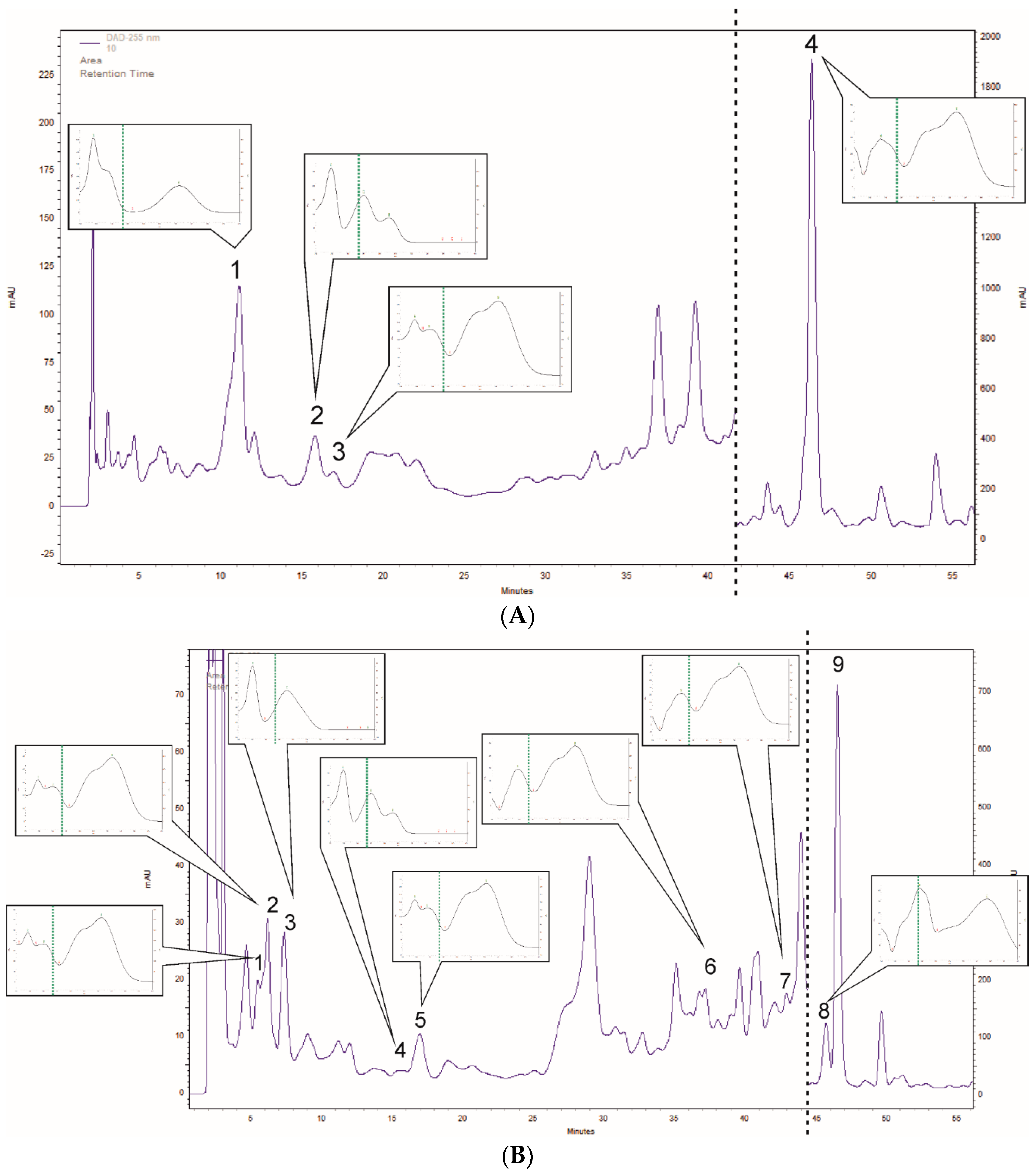
3.1. Polyphenol Profiling of Rosmarinus Officinalis and Ocimum Basilicum Leaf Extracts

In the R. officinalis methanolic leaf extract, the HPLC-DAD qualitative and quantitative analyses of selected phenolic compounds confirmed very high amount of rosmarinic acid (4040.00 mg/100 g DW (dry weight)). Moreover, other polyphenols, including genistic aicd (199.9 mg/100 g DW), vanilic acid (36.6 mg/100 g DW), and caffeic acid (27.6 mg/100 g DW) were determined (Figure 1A, Table 1).
In O. basilicum leaf extracts, a larger number of polyphenols were determined (8 phenolic acids and 1 flavonoid). In phenolic acids, the major one was rosmarinic acid (1128.5 mg/100 g DW), followed by 3,4-dihydroxyphenylacetic acid (312.3 mg/100 g DW), caftaric acid (60.3 mg/100 g DW), neochlorogenic acid (38.6 mg/100 g DW), isochlorogenic acid (30.0 mg/100 g DW), caffeic acid (17.6 mg/100 g DW), ferulic acid (14.4 mg/100 g DW), and a small amount of vanilic acid (1.1 mg/100 g DW) (Figure 1B, Table 1).

3.2. Antioxidant Effects

R. officinalis and O. basilicum leaf extracts, as well as major compounds, showed significant antioxidant effects by several methods (Table 2). R. officinalis showed high antioxidant effects compared to O. basilicum, as measured by all methods. Further, R. officinalis showed a comparable antioxidant effect to rosmarinic acid. The highest antioxidant values (lowest IC50) were found in 3,4-Dihydroxyphenylacetic acid and rosmarinic acid. Rutoside showed relatively low antioxidant effects as compared to other identified compounds.

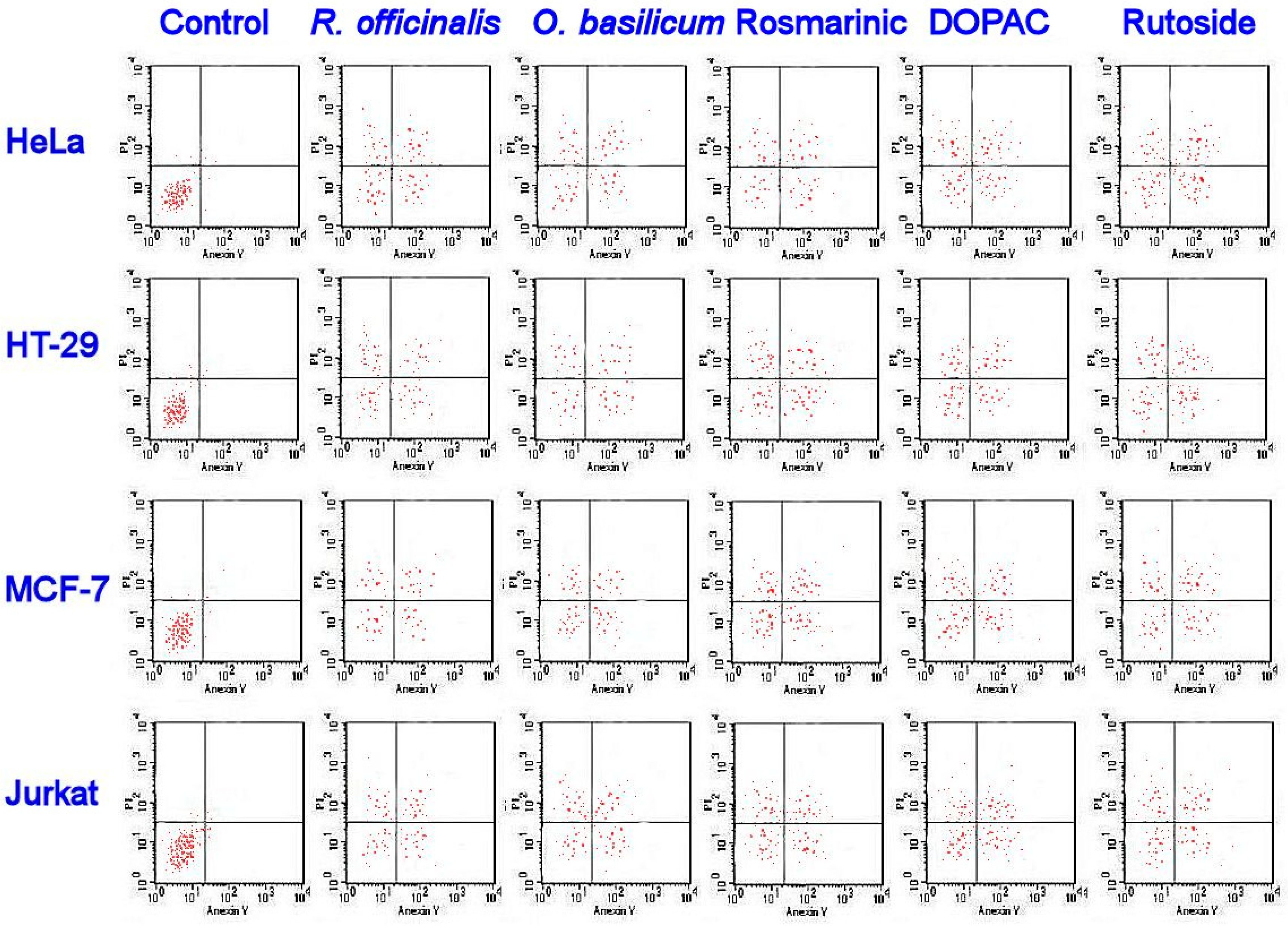
3.3. Antiproliferative and Cytotoxic Effects

The MTT assay of methanolic leaf extracts and identified phenols revealed obvious antiproliferative effects against T-cell lymphoblast like (Jurkat), breast adenocarcinoma cultures (MCF-7), colon adenocarcinoma (HT-29), cervical adenocarcinoma (HeLa), and HEK-293 (normal human cells), as shown in Table 3. The 3,4-Dihydroxyphenylacetic acid and rutoside had the lowest IC50 values compared to other compounds as well as leaf extracts.
The flow cytometry assay (Figure 2) showed the apoptotic cell accumulation the upper and lower right quadrant, following 48 h of exposure.

3.4. Antibacterial Effects

R. officinalis and O. basilicum methanolic leaf extracts had strong antibacterial effects against screened bacteria (Table 4). Staphylococcus aureus, Pseudomonas aeruginosa, and Micrococcus flavus were the most sensitive to leaf extracts. 3,4-dihydroxyphenylacetic acid, rutoside and caffeic acid were the most active isolated compounds against most bacteria. Gentisic acid was not active against all bacterial except Escherichia coli. Rosmarinic acid was weak against most bacteria and showed MBC > 500 mg mL−1.

3.5. Antifungal Activities

R. officinalis and O. basilicum methanolic leaf extracts showed antifungal activities against all fungi screened (Table 5). R. officinalis showed lower IC50 values than O. basilicum. 3,4-Dihydroxyphenylacetic acid, gentisic acid, rutoside, and caffeic acid showed strong antifungal activities against examined bacteria. Rosmarinic acid showed the weakest antifungal effects compared to other identified polyphenols.

4. Discussion

The rosmarinic acid in R. officinalis leaf extracts has been reported by several authors. However, the qualitative estimations are limited. Borrás-Linare et al. [43] studied 20 rosemary plants (harvested from different geographical zones of Serbia) by HPLC–ESI-QTOF-MS (high performance liquid chromatography coupled to electrospray quadrupole-time of flight mass spectrometry) method. The amount of rosmarinic acid varied from 50 to 2500 mg/100 g DW, depending on the place of harvest. Interestingly, the amount of rosmarinic acid quantified in their study was 1.6 times lower than that found in the North Saudi Arabia natural population origin samples in the current study. The rosmarinic acid was the unique phenolic acid confirmed by Kontogianni et al. [44] in the extracts of R. officinalis leaf commercial samples (Greece) using liquid chromatography/diode array detection/electrospray ion trap tandem mass spectrometry (LC/DAD/ESI-MS) method. The amount of rosmarinic acid from Greece [44] was equal to 1160 mg/100 g DW, which is 3.5 times lower than in the R. officinalis plants of Saudi origin of this study. The rosmarinic and caffeic acids in the R. officinalis leaf extracts samples obtained from United Kingdom ranged from 1000.0 to 1100.0 and from 10.0 to 20.0 mg/100 g DW, respectively [45]. These amounts were respectively c.a. 3.8 and 1.8 times lower than in the Saudi extracts of the present study. The vanillic acid was quantified (4 mg/100 g DW) by Luis et al. [46] in the leaf extracts of R. officinalis from United Kingdom. That amount was 9 times lower than in the samples of our collection. In addition, the same researchers detected rosmarinic acid (2150 mg/100 g DW) and caffeic acid (12 mg/100 g DW) in amounts respectively 1.9 and 2.3 times lower than the North Saudi Arabia natural population origin samples. Zgórka and Głowniak studied the compositions some phenolic acids in R. officinalis leaf extracts of Polish origin [47]. They detected relatively low amount of rosmarinic acid—c.a. 700 mg/100 g DW (5.8 times lower than in Saudi origin samples), caffeic acid—c.a. 25 mg/100 g DW (similar to Saudi origin samples) and vanillic acid—c.a. 15 mg/100 g DW (1.8 times lower Saudi origin samples). They also confirmed the gentisic acid in the extracts in amount 2 times lower than the current study samples (c.a. 50 mg/100 g DW).
In O. basilicum leaf extracts, the HPLC-DAD analyses revealed high amount of rosmarinic acid (1128.5 mg/100 g DW). This compound is generally recognized as typical for the Lamiaceae family species. For example, there are many studies describing the endogenous synthesis of high amounts of this compound of in vitro cultured biomass of O. basilicum of a different origin [48]. The phenolic composition of O. basilicum leaf extracts has not been studied deeply. One study investigated 16 different O. basilicum cultivars from North America [49]. There were considerable differences between tested cultivars and the main compound was rosmarinic acid (varied from 6 to 609 mg/100 g DW), followed by caftaric acid (from 9 to 49 mg/100 g DW) and caffeic acid (from 4 to 77 mg/100 g DW). These North American plants compositions of rosmarinic acid and caftaric acid were respectively 1.8 and 1.2 times lower than in the samples of Saudi origin, also the caffeic acid was 4.4 times higher. In O. basilicum plants from Iraq, the rosmarinic acid composition was equal to 306 mg/100 g DW, which is several times lower than Saudi one [50]. Zgórka and Głowniak [47] studied leaf extracts of Polish O. basilicum and they detected rosmarinic acid—c.a. 1100 mg/100 g DW, which is comparable to Saudi origin samples. Moreover, they confirmed also: caffeic acid—c.a. 270 mg/100 g DW (15 times more than in Ssaudi origin samples) and vanillic acid—c.a. 8 mg/100 g DW (7 times more than Saudi origin samples). The rosmarinic acid is the ester of caffeic acid with 3,4-dihydroxyphenylacetic acid (α-hydroxydihydrocaffeic acid). In the Saudi O. basilicum leaf extracts samples, we confirmed a high amount of 3,4-dihydroxyphenylacetic acid (α-hydroxydihydrocaffeic acid) (312.3 mg/100 g DW) and caffeic acid (17.6 mg/100 g DW). Vlase et al. studied the phenolic composition of O. basilicum leaf extracts of Romanian origin [51], and the major compound was rutoside (42.57 mg/100 g DW). The amount of rutoside was 3 times higher in the Saudi Samples than those of Romanian origin. In the Romanian samples, they also confirmed the availability of ferulic acid (0.7 mg/100 g DW), p-coumaric acid (2.1 mg/100 g DW), and trace amounts (below 0.02 mg/1000 g DW) of caffeic acid, caftaric acid, chlorogenic acid and rosmarinic acid. In the Saudi plants, we detected considerably high amounts of these compounds, especially for chlorogenic acid derivatives—caftaric acid (60.3 mg/100 g DW), neochlorogenic acid (38.6 mg/100 g DW)—as well as isochlorogenic acid (30.0 mg/100 g DW).
Leaf extracts of R. officinalis and O. basilicum showed antioxidant effects, as revealed by several methods. The antioxidant activity of Northern Riyadh R. officinalis leaf extracts is mainly attributed to major identified polyphenols including rosmarinic acid. The rosmarinic acid showed comparable antioxidant activities to R. officinalis leaf extracts. Previous investigation on R. officinalis from Morocco revealed antioxidant activities of ethanolic leaf extracts, and this was attributed to the major identified polyphenols, including carnosol and carnosic acid [52]. Other reports indicated that the major leaf extract polyphenols responsible for antioxidant activities might be rosmarinic acid, hesperidin, and rosmanol as well [53]. The rosmarinic acid itself has strong antioxidant activities, as found in this study, as well as in previous investigations [54]. Other compounds that were also present in the extracts of rosemary in this study, including gentisic acid, showed antioxidant activities and have been associated with antioxidant activities against gamma irradiation [25] and as an antioxidant agent food industries [32]. In O. basilicum, there were also antioxidant activities in leaf extracts, and this might be attributed to major identified polyphenols including rosmarinic acid, DOPAC and rutoside. DOPAC, has known antioxidant activities as revealed in previous studies [55]. Rutoside (rutin) is known also as potent antioxidant [56]. Previous studied revealed antioxidant activities of leaf extracts of O. basilicum [24,25,57].
Antiproliferative and cytotoxic activities against Jurkat, MCF-7, HT-29, and HeLa were found in the methanolic leaf extracts of R. officinalis in the current study. It was attributed to DOPAC and rosmarinic acid, as found in the MTT assay and flowcytometry. A previous report found that rosemary leaf extracts (from Germany) may have antiproliferative activities against human ovarian cancer A2780, and this activity might be attributed to main detected polyphenols such as carnosol, carnosic acid, and rosmarinic acid [58]. They described the perturbation of cell cycle progression in different phases of the cell cycle in response to leaf extract treatment. In the current study, there was an accumulation of necrotic and apoptotic cells in treated samples with the Saudi rosemary leaf extracts. The effects of rosmarinic acid and DOPAC on cancer cells had been described before. A previous investigation showed that rosemary leaf extracts had antiproliferative activities against HT-29 cancer cells by inhibiting cell proliferation, increasing cell cycle arrest, and increasing apoptosis [59]. The major composition of this extract was the rosmarinic acid [60]. The rosmarinic acid has strong anticancer activities against MCF-7 [61] and HT-29 [62]. The DOPAC has antiproliferative activities against few cancer cells, such as CHO-K1 [63]. In the current study, O. basilicum leaf extracts showed noticeable antiproliferative and cytotoxic activities against cancer cells, and this could be attributed to the polyphenolic composition, including rosmarinic acid, DOPAC, rutoside, ferulic acid, caffeic acid. Egyptian O. basilicum showed leaf methanolic extracts showed cytotoxic activities against HCT116 and HEBG2 cancer cells, and this was attributed to the phenolic composition, including caffeic acid, ferulic acid, catechin, quercetin, and others [64]. These polyphenols had known antiproliferative and cytotoxic activities, for example the rutoside obtained from Ferocactus species showed strong antiproliferative and cytotoxic activities against cancer cells [39].
The leaf extracts obtained from Saudi R. officinalis and O. basilicum had strong antibacterial and antifungal effects against screened bacteria and fungi. S. aureus, P. aeruginosa, and M. flavus were the most sensitive to leaf extracts. The antibacterial activity of leaf extracts is associated with some major polyphenols including DOPAC and rutoside. However, other polyphenols, including gentisic acid and rosmarinic acid were not relatively active. The antibacterial activity of DOPAC had been reported before against several bacteria [39]. Rutoside also has antibacterial activities against B. cereus and Salmonella enteritidis [65], S. aureus [66], and other bacteria [39]. R. officinalis and O. basilicum (from different regions) leaf extracts showed antibacterial activities in previous investigations [67]. Caffeic acid showed strong antibacterial activities, which is in agreement with previous studied confirming the antibacterial activities against different bacteria [5].
The antifungal activities of R. officinalis and O. basilicum methanolic leaf extracts had been reported before against several fungi [68]. However, this is the first report on the antifungal activities of R. officinalis from Northern Saudi Arabia. These antifungal activities are strongly associated with identified polyphenols, including 3,4-dihydroxyphenylacetic acid, gentisic acid, and rutoside. Gentisic acid is a dihydroxybenzoic acid that have known antifungal activities [69]. Similarly, DOPAC, rutoside, and caffeic acid had strong antifungal activities [5,39].

5. Conclusions

This is a novel report exploring the polyphenolic profile of natural populations of R. officinalis and O. basilicum in Northern Riyadh in Saudi Arabia. The HPLC-DAD analyses confirmed several polyphenols, including rosmarinic acid, gentisic acid, 3,4-dihydroxyphenylacetic acid (α-hydroxydihydrocaffeic acid), rutoside, and others out of 38 screened polyphenolic compounds. Rosmarinic acid was the major polyphenol in both studied species. R. officinalis methanolic leaf extracts contained rosmarinic acid and gentisic acid as major polyphenols, while O. basilicum contained rosmarinic acid, 3,4-dihydroxyphenylacetic acid, and rutoside, as well as others. R. officinalis showed higher antioxidant activities than O. basilicum using DPPH, FRAP, and β-carotene bleaching assay. These higher activities are associated with a higher composition of rosmarinic acid in leaf extracts. The antioxidant activities of O. basilicum were attributed to identified phenols of rosmarinic acid, 3,4-dihydroxyphenylacetic acid, and rutoside. There were antiproliferative and cytotoxic activities of leaf extracts, as well as identified polyphenols against several cancer cells. These activities were attributed to the accumulation of necrotic and apoptotic cells in treated cancer cells, with leaf extracts as well as identified polyphenols. The antibacterial and antifungal activities of leaf extracts were mainly attributed to 3,4-dihydroxyphenylacetic acid and rutoside in O. basilicum and rosmarinic acid and caffeic acid in R. officinalis. This study proved that R. officinalis and O. basilicum natural populations might be considered as promising sources of natural polyphenols with biological activities.

Author Contributions

Conceptualization, H.O.E., A.S., P.K., H.E., D.O.E.-A. and E.A.M.; Data curation, A.S. and E.A.M.; Formal analysis, H.O.E., A.S., P.K., H.E., D.O.E.-A., F.A.A.-M. and E.A.M.; Funding acquisition, H.O.E., H.E. and F.A.A.-M.; Investigation, H.O.E., A.S., P.K., H.E. and E.A.M.; Methodology, H.E., A.S. and D.O.E.-A.; Resources, P.K.; Software, A.S.; Validation, F.A.A.-M.; Visualization, P.K.; Writing–original draft, H.O.E., A.S., H.E., D.O.E.-A. and E.A.M.; Writing–review & editing, H.E. and F.A.A.-M. All authors have read and agreed to the published version of the manuscript.

Funding

The work was supported by King Saud University, Researchers Supporting Project number (RSP-2019/118).

Acknowledgments

The authors extend their appreciation to King Saud University, Researchers Supporting Project for funding this work through research group (RSP-2019/118).

Conflicts of Interest

The authors declare no conflict of interest.

References

  1. Halagarda, M.; Groth, S.; Popek, S.; Rohn, S.; Pedan, V. Antioxidant Activity and Phenolic Profile of Selected Organic and Conventional Honeys from Poland. Antioxidants 2020, 9, 44. [Google Scholar] [CrossRef] [PubMed] [Green Version]
  2. Okla, K.M.; Alamri, A.S.; Salem, Z.M.M.; Ali, M.H.; Behiry, I.S.; Nasser, A.R.; Alaraidh, A.I.; Al-Ghtani, M.S.; Soufan, W. Yield, Phytochemical Constituents, and Antibacterial Activity of Essential Oils from the Leaves/Twigs, Branches, Branch Wood, and Branch Bark of Sour Orange (Citrus aurantium L.). Processes 2019, 7, 363. [Google Scholar] [CrossRef] [Green Version]
  3. Salem, M.Z.M.; Elansary, H.O.; Ali, H.M.; El-Settawy, A.A.; Elshikh, M.S.; Abdel-Salam, E.M.; Skalicka-Wozniak, K. Bioactivity of essential oils extracted from Cupressus macrocarpa branchlets and Corymbia citriodora leaves grown in Egypt. BMC Complement. Altern. Med. 2018, 18, 23. [Google Scholar] [CrossRef] [PubMed]
  4. Elansary, H.O.; Szopa, A.; Kubica, P.; Al-Mana, F.A.; Mahmoud, E.A.; El-Abedin, T.K.A.Z.; Mattar, M.A.; Ekiert, H. Phenolic Compounds of Catalpa speciosa, Taxus cuspidata, and Magnolia acuminata have Antioxidant and Anticancer Activity. Molecules 2019, 24, 412. [Google Scholar] [CrossRef] [PubMed] [Green Version]
  5. Elansary, H.O.; Szopa, A.; Kubica, P.; Ekiert, H.; Mattar, M.A.; Al-Yafrasi, M.A.; El-Ansary, D.O.; Zin El-Abedin, T.K.; Yessoufou, K. Polyphenol Profile and Pharmaceutical Potential of Quercus spp. Bark Extracts. Plants 2019, 8, 486. [Google Scholar] [CrossRef] [PubMed] [Green Version]
  6. Elansary, H.O. Tree Bark Phenols Regulate the Physiological and Biochemical Performance of Gladiolus Flowers. Processes 2020, 8, 71. [Google Scholar] [CrossRef] [Green Version]
  7. Elansary, H.O.; Szopa, A.; Klimek-Szczykutowicz, M.; Jafernik, K.; Ekiert, H.; Mahmoud, E.A.; Barakat, A.A.; El-Ansary, D.O. Mammillaria Species—Polyphenols Studies and Anti-Cancer, Anti-Oxidant, and Anti-Bacterial Activities. Molecules 2019, 25, 131. [Google Scholar] [CrossRef] [Green Version]
  8. Khan, M.; Siddiqui, S.A. Concurrent chemoradiotherapy with or without induction chemotherapy for the management of cervical lymph node metastasis from unknown primary tumor. J. Cancer Res. Ther. 2018, 14, 1117–1120. [Google Scholar] [CrossRef]
  9. Elansary, H.O.M.; Adamec, L.; Štorchová, H. Uniformity of organellar DNA in Aldrovanda vesiculosa, an endangered aquatic carnivorous species, distributed across four continents. Aquat. Bot. 2010, 92, 214–220. [Google Scholar] [CrossRef]
  10. Alvarado-Sansininea, J.J.; Sánchez-Sánchez, L.; López-Muñoz, H.; Escobar, M.L.; Flores-Guzmán, F.; Tavera-Hernández, R.; Jiménez-Estrada, M. Quercetagetin and Patuletin: Antiproliferative, Necrotic and Apoptotic Activity in Tumor Cell Lines. Molecules 2018, 23, 2579. [Google Scholar] [CrossRef] [Green Version]
  11. Sezer, E.D.; Oktay, L.M.; Karadadaş, E.; Memmedov, H.; Selvi Gunel, N.; Sözmen, E. Assessing Anticancer Potential of Blueberry Flavonoids, Quercetin, Kaempferol, and Gentisic Acid, Through Oxidative Stress and Apoptosis Parameters on HCT-116 Cells. J. Med. Food 2019, 22, 1118–1126. [Google Scholar] [CrossRef] [PubMed]
  12. Flamini, G.; Cioni, P.L.; Morelli, I.; Macchia, M.; Ceccarini, L. Main Agronomic−Productive Characteristics of Two Ecotypes of Rosmarinus officinalis L. and Chemical Composition of Their Essential Oils. J. Agric. Food Chem. 2002, 50, 3512–3517. [Google Scholar] [CrossRef] [PubMed]
  13. Andrade, J.M.; Faustino, C.; Garcia, C.; Ladeiras, D.; Reis, C.P.; Rijo, P. Rosmarinus officinalis L.: An update review of its phytochemistry and biological activity. Future Sci. OA 2018, 4, FSO283. [Google Scholar] [CrossRef] [PubMed] [Green Version]
  14. EMA, C.O.H.M.P. Community Herbal Monograph on Rosmarinus Officinalis L., Folium, Doc. Ref.: EMEA/HMPC/13633/2009; ESCOP. 1997.; ‘Rosmarini folium’. Monographs on the Medicinal Uses of Plant Drug; European Scientific Cooperative on Phytotherapy: Exeter, UK, 2009. [Google Scholar]
  15. Council of Europe. European Pharmacopoeia 9.0; Council of Europe: Strasburg, France, 2017. [Google Scholar]
  16. U.S. Pharmacopeia. Food Chemicals Codex, 6th ed.; U.S. Pharmacopeia: Rockville, MD, USA, 2008; Available online: https://www.who.int/medicines/areas/quality_safety/quality_assurance/resources/US_Pharmacopoeia.pdf (accessed on 31 March 2020).
  17. Xhang, X. WHO Monographs on Selected Medicinal Plants WHO: Essential Medicines and Health Products Information Portal; World Health Organization: Geneva, Switzerland, 2004; Volume 2, p. 358. [Google Scholar]
  18. Elansary, H.O.; Mahmoud, E.A. Egyptian herbal tea infusions’ antioxidants and their antiproliferative and cytotoxic activities against cancer cells. Nat. Prod. Res. 2015, 29, 474–479. [Google Scholar] [CrossRef] [PubMed]
  19. Begum, A.; Sandhya, S.; Ali, S.; Ravindran, V.; Reddy, S.; Banji, D. An in-depth review on the medicinal flora Rosmarinus officinalis (Lamiaceae). Acta Sci. Pol. Technol. Aliment. 2013, 12, 61–74. [Google Scholar] [PubMed]
  20. Fahim, F.A.; Esmat, A.Y.; Fadel, H.M.; Hassan, K.F. Allied studies on the effect of Rosmarinus officinalis L. on experimental hepatotoxicity and mutagenesis. Int. J. Food Sci. Nutr. 1999, 50, 413–427. [Google Scholar] [CrossRef]
  21. Mangena, T.; Muyima, N.Y. Comparative evaluation of the antimicrobial activities of essential oils of Artemisia afra, Pteronia incana and Rosmarinus officinalis on selected bacteria and yeast strains. Lett. Appl. Microbiol. 1999, 28, 291–296. [Google Scholar] [CrossRef]
  22. Lis-Balchin, M.; Hart, S.; Deans, S.G.; Eaglesham, E. Comparison of the Pharmacological and Antimicrobial Action of Commercial Plant Essential Oils. J. Herbs Spices Med. Plants 1996, 4, 69–86. [Google Scholar] [CrossRef]
  23. El-Esawi, M.A.; Elansary, H.O.; El-Shanhorey, N.A.; Abdel-Hamid, A.M.E.; Ali, H.M.; Elshikh, M.S. Salicylic Acid-Regulated Antioxidant Mechanisms and Gene Expression Enhance Rosemary Performance under Saline Conditions. Front. Physiol. 2017, 8, 716. [Google Scholar] [CrossRef]
  24. Elansary, H.O.; Mahmoud, E.A. Basil cultivar identification using chemotyping still favored over genotyping using core barcodes and possible resources of antioxidants. J. Essent. Oil Res. 2015, 27, 82–87. [Google Scholar] [CrossRef]
  25. Elansary, H.O. Basil morphological and physiological performance under trinexapac-ethyl foliar sprays and prolonged irrigation intervals. Acta Physiol. Plant 2015, 37, 92. [Google Scholar] [CrossRef]
  26. Grayer, R.J.; Kite, G.C.; Goldstone, F.J.; Bryan, S.E.; Paton, A.; Putievsky, E. Infraspecific taxonomy and essential oil chemotypes in sweet basil, Ocimum basilicum. Phytochemistry 1996, 43, 1033–1039. [Google Scholar] [CrossRef]
  27. Hussain, A.; Anwar, F.; Sherazi, S.T.; Przybylski, R. Chemical Composition, Antioxidant and Antimicrobial Activities of Basil (Ocimum basilicum) Essential oils Depends on Seasonal Variations. Food Chem. 2008, 108, 986–995. [Google Scholar] [CrossRef] [PubMed]
  28. Da Silva, L.A.L.; Pezzini, B.R.; Soares, L. Spectrophotometric determination of the total flavonoid content in Ocimum basilicum L. (Lamiaceae) leaves. Pharmacogn. Mag. 2015, 11, 96–101. [Google Scholar] [PubMed] [Green Version]
  29. Flanigan, P.; Niemeyer, E. Effect of cultivar on phenolic levels, anthocyanin composition, and antioxidant properties in purple basil (Ocimum basilicum L.). Food Chem. 2014, 164, 518–526. [Google Scholar] [CrossRef] [PubMed]
  30. Bilal, A.; Jahan, N.; Makbul, S.; Bilal, S.N.; Habib, S.; Hajra, S. Phytochemical and pharmacological studies on Ocimum basilicum. Int. J. Curr. Res. Rev. 2012, 4, 73–83. [Google Scholar]
  31. Nayak, B. Phytochemical investigation and screening for anthelmintic activity of leafy extracts of various Ocimum (Tulsi) species. J. Pharm. Res. 2010, 3, 2140–2141. [Google Scholar]
  32. Elansary, H.O.; Szopa, A.; Kubica, P.; El-Ansary, D.O.; Ekiert, H.; Al-Mana, F.A. Malus baccata var. gracilis and Malus toringoides Bark Polyphenol Studies and Antioxidant, Antimicrobial and Anticancer Activities. Processes 2020, 8, 283. [Google Scholar] [CrossRef] [Green Version]
  33. Sulkowska-Ziaja, K.; Maslanka, A.; Szewczyk, A.; Muszynska, B. Physiologically Active Compounds in Four Species of Phellinus. Nat. Prod. Commun. 2017, 12, 363–366. [Google Scholar] [CrossRef] [Green Version]
  34. Szopa, A.; Kokotkiewicz, A.; Bednarz, M.; Luczkiewicz, M.; Ekiert, H. Studies on the accumulation of phenolic acids and flavonoids in different in vitro culture systems of Schisandra chinensis (Turcz.) Baill. using a DAD- HPLC method. Phytochem. Lett. 2017, 20, 462–469. [Google Scholar] [CrossRef]
  35. Yessoufou, K.; Elansary, H.O.; Mahmoud, E.A.; Skalicka-Wozniak, K. Antifungal, antibacterial and anticancer activities of Ficus drupacea L. stem bark extract and biologically active isolated compounds. Ind. Crops Prod. 2015, 74, 752–758. [Google Scholar] [CrossRef]
  36. Elansary, H.O.; Abdelgaleil, S.A.M.; Mahmoud, E.A.; Yessoufou, K.; Elhindi, K.; El-Hendawy, S. Effective antioxidant, antimicrobial and anticancer activities of essential oils of horticultural aromatic crops in northern Egypt. BMC Complement. Altern. Med. 2018, 18. [Google Scholar] [CrossRef] [PubMed]
  37. Elansary, H.O.; Yessoufou, K.; Abdel-Hamid, A.M.E.; El-Esawi, M.A.; Ali, H.M.; Elshikh, M.S. Seaweed Extracts Enhance Salam Turfgrass Performance during Prolonged Irrigation Intervals and Saline Shock. Front. Plant Sci. 2017, 8, 830. [Google Scholar] [CrossRef] [Green Version]
  38. El-Esawi, A.M.; Elkelish, A.; Soliman, M.; Elansary, O.H.; Zaid, A.; Shabir, W.H. Serratia marcescens BM1 Enhances Cadmium Stress Tolerance and Phytoremediation Potential of Soybean through Modulation of Osmolytes, Leaf Gas Exchange, Antioxidant Machinery, and Stress-Responsive Genes Expression. Antioxidants 2020, 9, 43. [Google Scholar] [CrossRef] [Green Version]
  39. Elansary, H.O.; Agnieszka, S.; Klimek-Szczykutowicz, M.; Ekiert, H.; Barakat, A.A.; Al-Mana, F.A. Antiproliferative, Antimicrobial, and Antifungal Activities of Polyphenol Extracts from Ferocactus Species. Processes 2020, 8, 138. [Google Scholar] [CrossRef] [Green Version]
  40. Elansary, H.O.; Szopa, A.; Kubica, P.; Ekiert, H.; Ali, H.M.; Elshikh, M.S.; Abdel-Salam, E.M.; El-Esawi, M.; El-Ansary, D.O. Bioactivities of Traditional Medicinal Plants in Alexandria. Evid. Based Complement. Altern. Med. 2018, 2018, 1463579. [Google Scholar] [CrossRef] [Green Version]
  41. Elansary, H.O.; Yessoufou, K.; Shokralla, S.; Mahmoud, E.A.; Skaicka-Wozniak, K. Enhancing mint and basil oil composition and antibacterial activity using seaweed extracts. Ind. Crops Prod. 2016, 92, 50–56. [Google Scholar] [CrossRef]
  42. Abd El-Kareem, M.S.M.; Mohamed, A.R.; Elansary, H.O.; Al-Mana, F.A. Mass Spectral Fragmentation of Pelargonium graveolens Essential Oil Using GC–MS Semi-Empirical Calculations and Biological Potential. Processes 2020, 8, 128. [Google Scholar] [CrossRef] [Green Version]
  43. Borrás-Linares, I.; Stojanovic, Z.; Quirantes-Piné, R.; Arráez-Román, D.; Švarc-Gajić, J.; Fernández-Gutiérrez, A.; Segura Carretero, A. Rosmarinus Officinalis Leaves as a Natural Source of Bioactive Compounds. Int. J. Mol. Sci. 2014, 15, 20585–20606. [Google Scholar] [CrossRef]
  44. Kontogianni, V.; Tomic, G.; Nikolic, I.; Nerantzaki, A.; Sayyad, N.; Stosic-Grujicic, S.; Stojanovic, I.; Gerothanassis, I.; Tzakos, A. Phytochemical profile of Rosmarinus officinalis and Salvia officinalis extracts and correlation to their antioxidant and anti-proliferative activity. Food Chem. 2013, 136, 120–129. [Google Scholar] [CrossRef]
  45. Wang, H.; Provan, G.; Helliwell, K. Determination of rosmarinic acid and caffeic acid in aromatic herbs by HPLC. Food Chem. 2004, 87, 307–311. [Google Scholar] [CrossRef]
  46. Luis, J.; Pérez, R.; Valdés, F. UV-B radiation effects on foliar concentrations of rosmarinic and carnosic acids in rosemary plants. Food Chem. 2007, 101, 1211–1215. [Google Scholar] [CrossRef]
  47. Zgórka, G.; Głowniak, K. Variation of free phenolic acids in medicinal plants belonging to the Lamiaceae family. J. Pharm. Biomed. Anal. 2001, 26, 79–87. [Google Scholar] [CrossRef]
  48. Kintzios, S.; Makri, O.; Panagiotopoulos, E.; Scapeti, M. In vitro rosmarinic acid accumulation in sweet basil (Ocimum basilicum L.). Biotechnol. Lett. 2003, 25, 405–408. [Google Scholar] [CrossRef] [PubMed]
  49. Kwee, E.; Niemeyer, E. Variations in phenolic composition and antioxidant properties among 15 basil (Ocimum basilicum L.) cultivars. Food Chem. 2011, 128, 1044–1050. [Google Scholar] [CrossRef]
  50. Adham, A.N. Comparative extraction methods, phytochemical constituents, fluorescence analysis and HPLC validation of rosmarinic acid content in Mentha piperita, Mentha longifolia and Osimum basilicum. J. Pharmacogn. Phytochem. 2015, 3, 130–139. [Google Scholar]
  51. Vlase, L.; Benedec, D.; Hanganu, D.; Damian, G.; Csillag, I.; Sevastre, B.; Mot, A.; Silaghi-Dumitrescu, R.; Tilea, I. Evaluation of Antioxidant and Antimicrobial Activities and Phenolic Profile for Hyssopus officinalis, Ocimum basilicum and Teucrium chamaedrys. Molecules 2014, 19, 5490–5507. [Google Scholar] [CrossRef]
  52. Bourhia, M.; Laasri, F.E.; Aourik, H.; Boukhris, A.; Ullah, R.; Bari, A.; Ali, S.S.; El Mzibri, M.; Benbacer, L.; Gmouh, S. Antioxidant and Antiproliferative Activities of Bioactive Compounds Contained in Rosmarinus officinalis Used in the Mediterranean Diet. Evid. Based Complement. Altern. Med. 2019, 2019. [Google Scholar] [CrossRef] [Green Version]
  53. Nieto, G.; Ros, G.; Castillo, J. Antioxidant and Antimicrobial Properties of Rosemary (Rosmarinus officinalis, L.): A Review. Medicines 2018, 5, 98. [Google Scholar] [CrossRef] [Green Version]
  54. Świsłocka, R.; Regulska, E.; Karpińska, J.; Świderski, G.; Lewandowski, W. Molecular Structure and Antioxidant Properties of Alkali Metal Salts of Rosmarinic Acid. Experimental and DFT Studies. Molecules 2019, 24, 2645. [Google Scholar] [CrossRef] [Green Version]
  55. Tang, Y.; Nakashima, S.; Saiki, S.; Myoi, Y.; Abe, N.; Kuwazuru, S.; Zhu, B.; Ashida, H.; Murata, Y.; Nakamura, Y. 3,4-Dihydroxyphenylacetic acid is a predominant biologically-active catabolite of quercetin glycosides. Food Res. Int. 2016, 89, 716–723. [Google Scholar] [CrossRef] [PubMed]
  56. Enogieru, A.B.; Haylett, W.; Hiss, D.C.; Bardien, S.; Ekpo, O.E. Rutin as a Potent Antioxidant: Implications for Neurodegenerative Disorders. Oxid. Med. Cell. Longev. 2018, 2018, 6241017. [Google Scholar] [CrossRef] [PubMed]
  57. Elansary, H.O.; Mahmoud, E.A. In vitro antioxidant and antiproliferative activities of six international basil cultivars. Nat. Prod. Res. 2015, 29, 2149–2154. [Google Scholar] [CrossRef] [PubMed]
  58. Tai, J.; Cheung, S.; Wu, M.; Hasman, D. Antiproliferation effect of Rosemary (Rosmarinus officinalis) on human ovarian cancer cells in vitro. Phytomedicine 2012, 19, 436–443. [Google Scholar] [CrossRef]
  59. Valdés, A.; Sullini, G.; Ibáñez, E.; Cifuentes, A.; García-Cañas, V. Rosemary polyphenols induce unfolded protein response and changes in cholesterol metabolism in colon cancer cells. J. Funct. Foods 2015, 15, 429–439. [Google Scholar] [CrossRef]
  60. Afonso, M.S.; de O Silva, A.M.; Carvalho, E.B.; Rivelli, D.P.; Barros, S.B.; Rogero, M.M.; Lottenberg, A.M.; Torres, R.P.; Mancini-Filho, J. Phenolic compounds from Rosemary (Rosmarinus officinalis L.) attenuate oxidative stress and reduce blood cholesterol concentrations in diet-induced hypercholesterolemic rats. Nutr. Metab. (Lond.) 2013, 10, 19. [Google Scholar] [CrossRef] [Green Version]
  61. Berdowska, I.; Zieliński, B.; Fecka, I.; Kulbacka, J.; Saczko, J.; Gamian, A. Cytotoxic impact of phenolics from Lamiaceae species on human breast cancer cells. Food Chem. 2013, 141, 1313–1321. [Google Scholar] [CrossRef]
  62. Scheckel, K.A.; Degner, S.C.; Romagnolo, D.F. Rosmarinic acid antagonizes activator protein-1-dependent activation of cyclooxygenase-2 expression in human cancer and nonmalignant cell lines. J. Nutr. 2008, 138, 2098–2105. [Google Scholar] [CrossRef]
  63. Maggio, R.; Armogida, M.; Scarselli, M.; Salvadori, F.; Longoni, B.; Pardini, C.; Chiarenza, A.; Chiacchio, S.; Vaglini, F.; Bernardini, R.; et al. Dopamine agonists and analogues have an antiproliferative effect on CHO-K1 cells. Neurotox. Res. 1999, 1, 285–297. [Google Scholar] [CrossRef]
  64. Abd El Azim, M.; Abdelgawad, A.; MohamedEl, G.; Ali, S.; el-Mousallami, A. Phenolic Compounds and Cytotoxic Activities of Methanol Extract of Basil (Ocimum basilicum L.). J. Microb. Biochem. Technol. 2015, 7, 185. [Google Scholar] [CrossRef]
  65. Arima, H.; Ashida, H.; Danno, G.-I. Rutin-enhanced Antibacterial Activities of Flavonoids against Bacillus cereus and Salmonella enteritidis. Biosci. Biotechnol. Biochem. 2002, 66, 1009–1014. [Google Scholar] [CrossRef] [PubMed] [Green Version]
  66. Amin, M.U.; Khurram, M.; Khattak, B.; Khan, J. Antibiotic additive and synergistic action of rutin, morin and quercetin against methicillin resistant Staphylococcus aureus. BMC Complement. Altern. Med. 2015, 15, 59. [Google Scholar] [CrossRef] [PubMed] [Green Version]
  67. Genena, A.K.; Hense, H.; Smânia Junior, A.; Souza, S.M.D. Rosemary (Rosmarinus officinalis): A study of the composition, antioxidant and antimicrobial activities of extracts obtained with supercritical carbon dioxide. Food Sci. Technol. 2008, 28, 463–469. [Google Scholar] [CrossRef] [Green Version]
  68. Nugroho, C.; Mirnia, E.; Cumagun, C.J. Antifungal Activities of Sweet Basil (Ocimum basilicum L.) Aqueous Extract against Sclerotium rolfsii, Causal Agent of Damping-Off on Tomato Seedling. AGRIVITA J. Agric. Sci. 2019, 41. [Google Scholar] [CrossRef]
  69. Pj, B.; Shibumon, G.; Sunny, K.; George, S. 2, 3-Dihydroxybenzoic Acid: An Effective Antifungal Agent Isolated from Flacourtia inermis Fruit. Int. J. Pharm. Clin. Res. 2010, 2, 101–105. [Google Scholar]
Figure 1. The examples of HPLC-DAD chromatographic separation (λ = 254 nm) of leaf extracts of: (A) R. officinalis (1—gentisic acid, 2—vanillic acid, 3—caffeic acid, 4—rosmarinic acid); and (B) O. basilicum (1—neochlorogenic acid, 2—caftaric acid, 3—3,4-dihydroxyphenylacetic acid, 4—vanillic acid, 5—caffeic acid, 6—ferulic acid, 7—isochlorogenic acid, 8—rutoside, 9—rosmarinic acid).
Processes 08 00446 g001
Figure 2. Cytotoxicity of R. officinalis and O. basilicum leaf extracts, and rosmarinic acid, 3,4-dihydroxyphenylacetic acid (DOPAC), and rutoside, as estimated with flow cytometry. There was an accumulation of apoptotic cells in the early (lower right quadrant) and late apoptotic (upper right quadrant).
Processes 08 00446 g002
Table 1. The contents of polyphenols in R. officinalis and O. basilicum leaf extracts (mg/100g DW ± SD).
CompoundRosmarinus officinalisOcimum basilicum
Caffeic acid27.6 ± 3.617.6 ± 1.3
Caftaric acidnd *60.3 ± 4.5
3,4-Dihydroxyphenylacetic acidnd312.3 ± 20.4
Ferulic acidnd14.4 ± 1.7
Gentisic acid119.9 ± 2.0nd
Isochlorogenic acidnd30.0 ± 3.3
Neochlorogenic acidnd38.6 ± 2.7
Rosmarinic acid4040.0 ± 189.21128.5 ± 70.4
Vanillic acid36.6 ± 1.71.1 ± 0.0
Rutosidend139.4 ± 18.8
* nd—not detected.
Table 2. 2,2-diphenyl-1-picrylhydrazyl (DPPH), β-Carotene bleaching and ferric reducing antioxidant power (FRAP) measures of R. officinalis and O. basilicum leaf extracts, rosmarininic acid, 3,4-dihydroxyphenylacetic acid, gentisic acid, and rutoside.
DPPH (IC50, µg mL−1)β-Carotene-Bleaching Assay (IC50, µg mL−1)FRAP (IC50, mM TEAC/g Extract)
R. officinalis2.9 ± 0.1c3.6 ± 0.1c4.1 ± 0.2e
O. basilicum4.7 ± 0.1cd5.1 ± 0.3d5.5 ± 0.1e
Rosmarinic acid 2.6 ± 0.1d3.0 ± 0.1d3.3 ± 0.9f
3,4-Dihydroxyphenylacetic acid 2.1 ± 0.1d2.7 ± 0.1d3.2 ± 0.1d
Gentisic acid 5.8 ± 0.1d7.3 ± 0.2d9.5 ± 1.0f
Rutoside 14.3 ± 1.1d16.5 ± 0.7d18.6 ± 0.9d
BHT2.6 ± 0.2e3.1 ± 0.1e-
Trolox--3.1 ± 0.1g
Different letters within a column indicates significant differences (p ≤ 0.05). TEAC: Trolox equivalents antioxidant.
Table 3. Antiproliferative activity (IC50 (µg mL−1)) of R. officinalis and O. basilicum leaf extracts, rosmarinic acid, 3,4-dihydroxyphenylacetic acid, gentisic acid, and rutoside on cancer cells.
HeLaHT-29MCF-7JurkatHEK-293
R. officinalis39.52 ± 2.336.59 ± 2.738.29 ± 1.764.39 ± 3.1˃400
O. basilicum45.26 ± 3.248.31 ± 2.947.23 ± 3.175.13 ± 3.6˃400
Rosmarinic acid36.30 ± 1.324.27 ± 2.125.61 ± 2.547.43 ± 3.1˃400
3,4-Dihydroxyphenylacetic acid3.11 ± 0.1f20.41 ± 0.77.12 ± 0.95.63 ± 1.1˃400
Gentisic acid5.7 ± 0.524.26 ± 0.910.13 ± 0.77.4 ± 0.6˃400
Rutoside3.9 ± 0117.5 ± 0.8f5.0 ± 0.3f4.0 ± 0.5˃400
Vinblastine sulfate2.1 ± 0.04 16.3 ± 0.70.11 ± 0.0142.7 ± 1.3
Taxol0.07 ± 0.004
Table 4. R. officinalis and O. basilicum leaf extracts, rosmarinic acid, 3,4-dihydroxyphenylacetic acid, gentisic acid, and rutoside antibacterial activities by means of minimum inhibitory (MIC) and bactericidal concentration (MBC) in (mg mL−1).
B. cereusP. aeruginosaL. monocytogenesE. coliM. flavusS. aureus
MICMICMICMICMICMIC
MBCMBCMBCMBCMBCMBC
R. officinalis0.56 ± 0.030.43 ± 0.030.54 ± 0.030.46 ± 0.020.36 ± 0.030.29 ± 0.03
0.97 ± 0.050.85 ± 0.030.96 ± 0.050.91 ± 0.030.82 ± 0.030.79 ± 0.02
O. basilicum0.43 ± 0.030.32 ± 0.020.41 ± 0.010.35 ± 0.010.27 ± 0.020.23 ± 0.01
0.85 ± 0.050.75 ± 0.030.89 ± 0.030.68 ± 0.030.59 ± 0.030.56 ± 0.03
Rosmarinic acid39.31 ± 1.1337.2 ± 1.6446.21 ± 3.1242.1 ± 3.4231.11 ± 1.5323.31 ± 0.96
>500>500>500>500>500>500
Gentisic acidN.D.N.D.N.D.23.00 ± 0.02N.D.N.D.
N.D.N.D.N.D.>100N.D.N.D.
3,4-Dihydroxyphenylacetic acid0.07 ± 0.010.06 ± 0.010.07 ± 0.010.08 ± 0.010.14 ± 0.020.13 ± 0.01
0.15 ± 0.010.13 ± 0.010.17 ± 0.010.19 ± 0.020.30 ± 0.020.27 ± 0.02
Rutoside0.10 ± 0.010.06 ± 0.010.10 ± 0.010.10 ± 0.010.13 ± 0.010.12 ± 0.01
0.20 ± 0.010.11 ± 0.010.20 ± 0.020.24 ± 0.010.24 ± 0.020.24 ± 0.02
Caffeic acid0.12 ± 0.010.06 ± 0.010.25 ± 0.010.10 ± 0.010.12 ± 0.010.21 ± 0.01
0.29 ± 0.030.12 ± 0.010.55 ± 0.030.23 ± 0.020.29 ± 0.020.43 ± 0.03
Streptomycin0.07 ± 0.010.09 ± 0.010.10 ± 0.010.10 ± 0.010.10 ± 0.010.15 ± 0.01
0.16 ± 0.010.19 ± 0.010.23 ± 0.010.21 ± 0.010.21 ± 0.010.31 ± 0.01
Table 5. Minimum inhibitory (MIC) and fungicidal concentration (MFC) of R. officinalis and O. basilicum leaf extracts, rosmarinic acid, 3,4-dihydroxyphenylacetic acid, gentisic acid and rutoside. Values are means of 3 replicates in (mg mL−1).
Aspergillus flavusAspergillus ochraceusAspergillus nigerCandida albicansPenicillium funiculosumPenicillium ochrochloron
MICMICMICMICMICMIC
MFCMFCMFCMFCMFCMFC
R. officinalis0.49 ± 0.050.56 ± 0.030.69 ± 0.030.86 ± 0.030.42 ± 0.030.49 ± 0.05
0.96 ± 0.051.18 ± 0.131.23 ± 0.071.59 ± 0.150.87 ± 0.050.93 ± 0.05
O. basilicum0.31 ± 0.020.38 ± 0.050.54 ± 0.050.63 ± 0.090.35 ± 0.030.38 ± 0.01
0.74 ± 0.030.89 ± 0.031.01 ± 0.091.16 ± 0.080.76 ± 0.050.89 ± 0.03
Rosmarinic acid181.92 ± 8.47222.32 ± 9.18231.65 ± 8.69385.7 ± 15.76218.86 ± 11.78349.9 ± 11.45
>1000>1000>1000>1000>1000>1000
Gentisic acid0.20 ± 0.030.15 ± 0.010.24 ± 0.030.29 ± 0.030.23 ± 0.010.18 ± 0.02
0.53 ± 0.050.49 ± 0.030.58 ± 0.040.65 ± 0.050.52 ± 0.030.43 ± 0.04
3,4-Dihydroxyphenylacetic acid0.19 ± 0.010.21 ± 0.020.25 ± 0.020.28 ± 0.030.27 ± 0.030.21 ± 0.01
0.41 ± 0.030.45 ± 0.030.51 ± 0.040.63 ± 0.050.63 ± 0.040.45 ± 0.05
Rutoside0.20 ± 0.020.19 ± 0.010.29 ± 0.030.24 ± 0.030.31 ± 0.020.21 ± 0.03
0.45 ± 0.030.56 ± 0.030.64 ± 0.050.53 ± 0.030.73 ± 0.040.47 ± 0.05
Caffeic acid0.21± 0.020.23 ± 0.010.20 ± 0.020.31 ± 0.010.26 ± 0.010.20 ± 0.01
0.43 ± 0.030.46 ± 0.030.40 ± 0.030.63 ± 0.030.61 ± 0.040.42 ± 0.03
KTZ (Ketoconazolum)0.22 ± 0.020.20 ± 0.010.10± 0.010.22 ± 0.012.00 ± 0.110.22 ± 0.01
0.45 ± 0.030.41 ± 0.030.21 ± 0.020.43 ± 0.033.54 ± 0.100.44 ± 0.03

