Article

Polyphenol Profile and Antimicrobial and Cytotoxic Activities of Natural Mentha × piperita and Mentha longifolia Populations in Northern Saudi Arabia

by
Hosam O. Elansary
1,2,3,*,
Agnieszka Szopa
4,*,
Paweł Kubica
4,
Halina Ekiert
4,
Marta Klimek-Szczykutowicz
4,
Diaa O. El-Ansary
5 and
Eman A. Mahmoud
6
1
Plant Production Department, College of Food and Agricultural Sciences, King Saud University, P.O. Box 2455, Riyadh 11451, Saudi Arabia
2
Floriculture, Ornamental Horticulture, and Garden Design Department, Faculty of Agriculture (El-Shatby), Alexandria University, Alexandria 21545, Egypt
3
Department of Geography, Environmental Management, and Energy Studies, University of Johannesburg, APK campus, Johannesburg 2006, South Africa
4
Department of Pharmaceutical Botany, Medical College, Jagiellonian University, ul. Medyczna 9, 30-688 Kraków, Poland
5
Precision Agriculture Laboratory, Department of Pomology, Faculty of Agriculture (El-Shatby), Alexandria University, Alexandria 21545, Egypt
6
Department of Food Industries, Faculty of Agriculture, Damietta University, Damietta 34517, Egypt
*
Authors to whom correspondence should be addressed.
Submission received: 25 March 2020 / Revised: 14 April 2020 / Accepted: 16 April 2020 / Published: 20 April 2020
(This article belongs to the Section Biological Processes and Systems)

Abstract

:
New sources of polyphenols with anticancer, antioxidant, and antimicrobial properties in arid environments are critical for the development of alternative medicines and natural remedies. This study explored the polyphenol profiles and biological activities of methanolic leaf extracts from natural Mentha × piperita and Mentha longifolia populations in northern Saudi Arabia. Chromatographic analyses identified several polyphenols in M. × piperita including phenolic acids: rosmarinic acid (1547.6 mg/100 g DW (dry weight)), cryptochlorogenic acid (91.7 mg/100 g DW), and chlorogenic acid (69.4 mg/100 g DW), as well as flavonoids: naringin (328.8 mg/100 g DW) and cynaroside (162.8 mg/100 g DW). The major polyphenols in M. longifolia were: rosmarinic acid (781.6 mg/100 g DW), cryptochlorogenic acid (191.1 mg/100 g DW), p-coumaric acid (113.0 mg/100 g DW), m-coumaric acid (112.2 mg/100 g DW), and chlorogenic acid (63.8 mg/100 g DW). M. × piperita and M. longifolia leaf extracts had high antioxidant activities due to the major polyphenols (cynaroside, rosmarinic and cryptochlorogenic acids). M. × piperita had higher activities against different cancer cells than M. longifolia. Naringin, cryptochlorogenic acid, and rosmarinic acid had the highest activities against cancer cells. The leaf extracts had antibacterial effects against most bacteria species (Pseudomonas aeruginosa was most sensitive), which was attributed to the polyphenols. Antifungal activities were similarly broad (Aspergillus flavus was most sensitive) and attributed to naringin, cryptochlorogenic acid, and caffeic acid. Populations of M. × piperita and M. longifolia in Northern Riyadh may be a valuable source of natural biologically active compounds.

1. Introduction

Polyphenols may be the most important secondary metabolite in the plant kingdom due to their pharmacological and medicinal properties. The medical applications of polyphenols include cancer control [1,2,3,4], bacterial and fungal infections [5,6], age elongation as antioxidants [7], and the treatment of diseases related to obesity [8], neurology [9], diabetes [10], and cardiovascular inflammation [11,12]. Polyphenols are also used in food processing and as functional foods [13].
The biochemical effects of polyphenols as antioxidants is based on their ability to scavenge free radicals and control metal chelation reactions [1,14,15,16,17,18,19]. In humans, healthy metabolic functioning is maintained by specific polyphenols that remove reactive oxygen species (ROS) such as hydrogen peroxide and singlet oxygen from the cells [7]. The anticancer properties of polyphenols are also partially related to antioxidant mechanisms, including the detoxification of enzymes and the reduction of free radicals [1,14,15,16,17,18,19]. The antiproliferative and apoptotic activities of polyphenols on cancer cells may be the backbone of the anticancer effects of dozens of polyphenols, via their arrestment of the cell cycle and molecular regulation of specific gene expression [1,14,15,16,17,18].
Bacterial and fungal infections cause serious human diseases such as pneumonia, soft tissue infection, sepsis, listeriosis, candidiasis, and peritonitis [20,21,22,23,24]. However, antibiotic resistance is an increasingly prevalent issue in the medical and scientific communities [25], and thus the discovery of new polyphenol sources to control these diseases is important. Medicinal plants are a key source of polyphenols—they have essential oils in the leaves and the leaf extracts contain phenolic acids, flavonoids, and catechins [26,27,28,29,30]. The Mentha genus in the Lamiaceae family contains several medicinal plants such as basil and sage, and Mentha piperita L. (syn. Mentha balsamea Wild.) is one of the most widely used herbal medicines [31].
Mentha piperita folium and Mentha piperita aetheroleum are listed in official phytotherapy documents, e.g., the World Health Organization (WHO) monographs on selected medicinal plants [32] in the European Pharmacopoeia [33]. Peppermint tea, brewed from the plant leaves, and peppermint essential oil are used as traditional medicines. Mentha longifolia L. (syn. M. spicata var. longifolia L., M. sylvestris L., M. tomentosa D’Urv, M. incana Willd.) is native to continental Europe, western and central Asia, and northern and southern (but not tropical) Africa. It is a variable herbaceous perennial with numerous morphological and phytochemical similarities to M. × piperita [34,35]. The Mentha genus is highly diverse, with more than 19 species, 13 hybrids, and new cultivars, all of which possess variable molecular, morphological, biochemical, and geographical properties [30,36,37,38,39]. The chemical composition of Mentha plants is influenced by many factors (e.g., geography, region, climate, or plant growth conditions), but the phytochemical composition is less variable among species [40]. Most research has focused on M. × piperita, revealing dozens of terpenes in the essential oil that is commonly used as a cough treatment or as an ointment [40]. The biological activity of Mentha has been attributed to the volatile oil constitutes, but the phenolic compounds may also play an important role [41]. The phenolic acid content is estimated to be 2.7–5.5%, consisting mainly of rosmarinic acid, caffeic acid, and protocatechuic acid. The flavonoid content is estimated to be 3.0–6.3% [42], containing mainly luteolin-7-O-rutinoside, eriocitrin, narirutin, and hesperidin [40,41].
In this study, natural M. × piperita and M. longifolia populations from northern Saudi Arabia were investigated for the chemical compositions and biological activities of their leaves methanolic extracts. The polyphenol profiles were qualitatively and quantitatively evaluated by high-performance liquid chromatography with diode-array detection (HPLC–DAD). The results presented here represent the first examination of the antioxidant, antiproliferative, cytotoxic effects, and the antibacterial and antifungal potential of these plants.

2. Materials and Methods

2.1. Plant Material and Preparation

Leaf extracts were obtained from natural M. × piperita and M. longifolia populations growing in the Riyadh region of northern Saudi Arabia. Plant samples were identified and vouchered at the College of Food and Agricultural Sciences, King Saud University (Hosam0002164–100). The leaves were dried in a lyophilizer, powdered, and then extracted with methanol (0.2 g DW (dry weight) in 10 mL) by sonication (Sonic-2, Polsonic) twice for 30 min at 30 °C. The extracts were purified with Whatman paper, and the residues were left to dry at room temperature (i.e., allowing methanol evaporation). The residues were stored at −80 °C. For liquid chromatography, the residues were dissolved in methanol (1 mL, Merck, Darmstadt, Germany), and for the bioassays, a rotary evaporator was used to remove methanol from the samples [2,43]. All of the experimental procedures were approved by the animal committee of the University of Johannesburg in collaboration with the Egyptian partners of Damietta (2018-2019-75364). The microorganisms and cancer cell lines (ATCC collection) were obtained from the Faculty of Agriculture, Alexandria, Egypt.

2.2. Analyses of Phenolic Compounds

The extracts were subjected to high-performance liquid chromatography with diode-array detection (HPLC –DAD) analyses on a Merck-Hitachi liquid chromatograph (LaChrom Elite, Merck, Darmstadt, Germany) equipped with a DAD detector (L-2455). Analyses were performed with the Purospher RP-18e column (250 × 4 mm; 5 μm, Merck, Darmstadt, Germany). The mobile phase was as follows: A—methanol, B—methanol: 0.5% acetic acid 1:4 (v/v), in a gradient of 100% B for 0–20 min; 100–80% B for 20–35 min; 80–60% B for 35–55 min; 60–0% B for 55–70 min; 0% B for 70–75 min; 0–100% B for 75–80 min; 100% B for 80–90 min. The temperature was 25 °C, the flow rate was 1 mL/min, and the injection volume was 20 µL. The compounds of interest were screened at 210 to 400 nm and quantitatively detected at 254 nm. This HPLC method was previously validated by our group [44,45]. The compounds were identified by comparison to the UV spectra and retention times of the reference substances and by cochromatography. Quantification was calculated from the calibration curves. A collection of commercially available phenolic acid compound standards (n = 26) were used for the qualification and quantification analyses. The standards included cinnamic acid and its derivatives (caffeic acid, o-coumaric acid, m-coumaric acid, p-coumaric acid, ferulic acid, hydrocaffeic acid, isoferulic acid, and sinapic acid), benzoic acid and its derivatives (3,4-dihydroxyphenylacetic acid, ellagic acid, gallic acid, gentisic acid, p-hydroxybenzoic acid, protocatechuic acid, salicylic acid, syringic acid, and vanillic acid), and depsides (caftaric acid, chlorogenic acid, isochlorogenic acid, cryptochlorogenic acid, neochlorogenic acid, and rosmarinic acid). The flavonoid standards (n = 16) included aglycones (cynaroside, myricetin, naringin, quercetin, kaempferol, rhamnetin, and luteolin) and glycosides (apigetrin, cynaroside, hyperoside, quercitrin, robinin, rutoside, isoquercetin, trifolin, and vitexin). All of the chemicals were acquired from Sigma-Aldrich (Berlin, Germany).

2.3. Antiproliferative and Cytotoxic Activities

The antiproliferative and cytotoxic activities of the leaf extracts were examined in T-cell lymphoblast-like (Jurkat), breast adenocarcinoma (MCF-7), colon adenocarcinoma (HT-29), HEK-293 (normal human cells), and cervical adenocarcinoma (HeLa) cell lines [1,2,46]. To measure the changes in cells viability, the MTT assay was used. Leaf extracts were solubilized in DMSO (1%). Cancer cells were grown in standard media (MEM) using (10% fetal bovine serum (FBS), 0.1 mM nonessential amino acids, 17.8 mM sodium bicarbonate (NaHCO3), and 1 mM sodium pyruvate in 75 cm2 flasks. The cells were seeded on microtiter plates at 4 × 10−4 cells per µL in 270 µL medium for 48 h (37 °C, 5% CO2). After 48 h, the extracts were added to final concentrations of 50, 100, 200, 300, and 400 µg/mL. The plates were washed with phosphate-buffered saline (PBS), and then 12 mM of MTT (dissolved in PBS) was added to the medium. Next, isopropanol (0.04 N hydrochloric acid, HCl) was added to each well, and the plates were left undisturbed for 40 min. Negative (untreated) and positive controls (vinblastine sulfate and taxol) were used, and the inhibition activity percentage (IAA) was calculated from the absorbance at 570 nm as:
I A A = ( A B 570 n m ) C ( A B 570 n m ) s ( A B 570 n m ) C × 100
where ( A B 570 n m ) C and ( A B 570 n m ) s are the absorbances (AB) at 570 nm for the control and test samples.
To determine the IC50, the percentage of viable cells was plotted against extract concentration in µg/mL. The IC50 was used in the flow cytometry assay to study the cytotoxic activities of leaf extracts and apoptotic cell populations (FAC Scan, USA) [1,46,47]. Cultured cancer cells in 6-well plates were subjected the IC50 of leaf extracts as well as naringin, cryptochlorogenic acid and rosmarinic acid. Untreated cells were considered as control for 48 h. Trypsin (0.25%) was used to detach cells in Hank’s balanced salt solution (Thermo Fisher Scientific, Berlin, Germany). The cells were stained using the Annexin V apoptosis detection kit (Sigma, St. Louis, MO, USA) by incubation in the dark at 37 °C for 15 min then washing with PBS. The data of flow cytometer is presented in quadrants: Lower left (viable cells), upper left (necrotic cells), lower right (early apoptotic cells), and upper right (late apoptotic cells).

2.4. Antioxidant Activity

The leaf extracts were examined by β-carotene bleaching, ferric reducing antioxidant power (FRAP), and DPPH (2,2-Diphenyl-1-picrylhydrazyl) assays [2,48,49,50,51,52]. The amount of leaf extract required to scavenge 50% of both solutions from the β-carotene bleaching and DPPH assays was named the IC50 (μg/mL). The IC50 value was determined by plotting the inhibition percent against extract concentration. The absorbance was measured at 470 nm for β-carotene bleaching, at 517 nm for DPPH, and at 593 nm for FRAP. The FRAP reagent was prepared as in previous work [43]. Aliquots (100 μL) of the leaf extracts/Trolox (Sigma-Aldrich, Berlin, Germany) were added to the reagent (3 mL), mixed, and incubated for 30 min at 37 °C. The FRAP calibration procedure involved serial dilutions of Trolox (0–0.5 Mmol/L) as the standard. Butylated hydroxytoluene (BHT) was used as the standard for β-carotene bleaching and DPPH. All of the assays were conducted in triplicate and repeated twice.

2.5. Antibacterial Effect

We assayed bacterial isolates of Listeria monocytogenes (clinical isolate), Staphylococcus aureus (ATCC 6538), Escherichia coli (ATCC 35210) ria, Bacillus cereus (ATCC 14579), Pseudomonas aeruginosa (ATCC 27853), and Micrococcus flavus (ATCC 10240). A microtiter plates-based protocol (micro-dilution) [4,23,29,53] was followed by mixing serial concentrations of the leaf extracts with bacterial inoculum (1.0 × 10⁴ CFU) and 100 μL of tryptic soy broth in each well. The plates were incubated for one day at 37 °C on a rotary shaker. The minimum inhibitory concentration (MIC) was defined as the lowest concentration that caused no visible growth. The minimum bacterial concentration (MBC) was determined by serial subculturing of the extracts (2 μL) and defined as the minimal concentration that caused 99.5% elimination of the inoculum. The optical density (OD) was measured at 655 nm, and a positive control (streptomycin; 0.01–10 mg/mL) was used alongside a negative control (DMSO, 1%).

2.6. Antifungal Effect

The antifungal effects of M. × piperita and M. longifolia were assayed for Penicillium ochrochloron (ATCC 48663), Aspergillus ochraceus (ATCC 12066), Candida albicans (ATCC 12066), Aspergillus niger (ATCC 6275), Aspergillus flavus (ATCC 9643), and Penicillium funiculosum (ATCC 56755) using a micro-dilution method [23,29,47]. The MIC was determined by stereomicroscope, and the minimum fungicidal concentration (MFC) was determined by preparing serial dilutions of the extracts (2 µL) in subcultures of fungi at 28 °C for 72 h in microtiter plates containing 100 µL of broth medium. Ketoconazole (KTZ, 1–3500 µg/mL) was used as a positive control and DMSO (1%) was used as a negative control.

2.7. Statistical Analyses

Differences among the groups were tested as the least significant differences (LSD) in SPSS software. The mean and standard deviation (SD) were calculated from the three replicates (of each assay).

3. Results

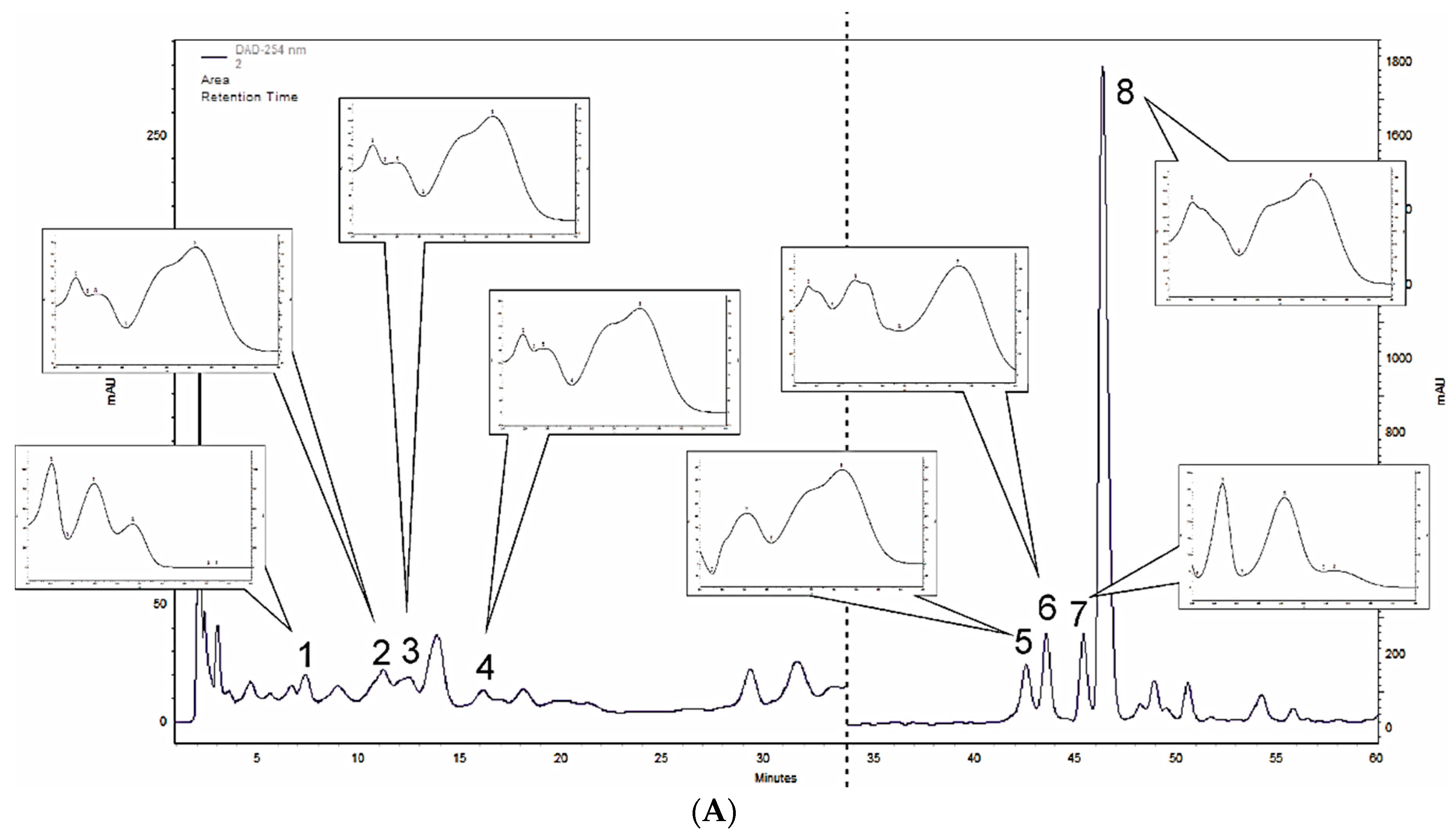
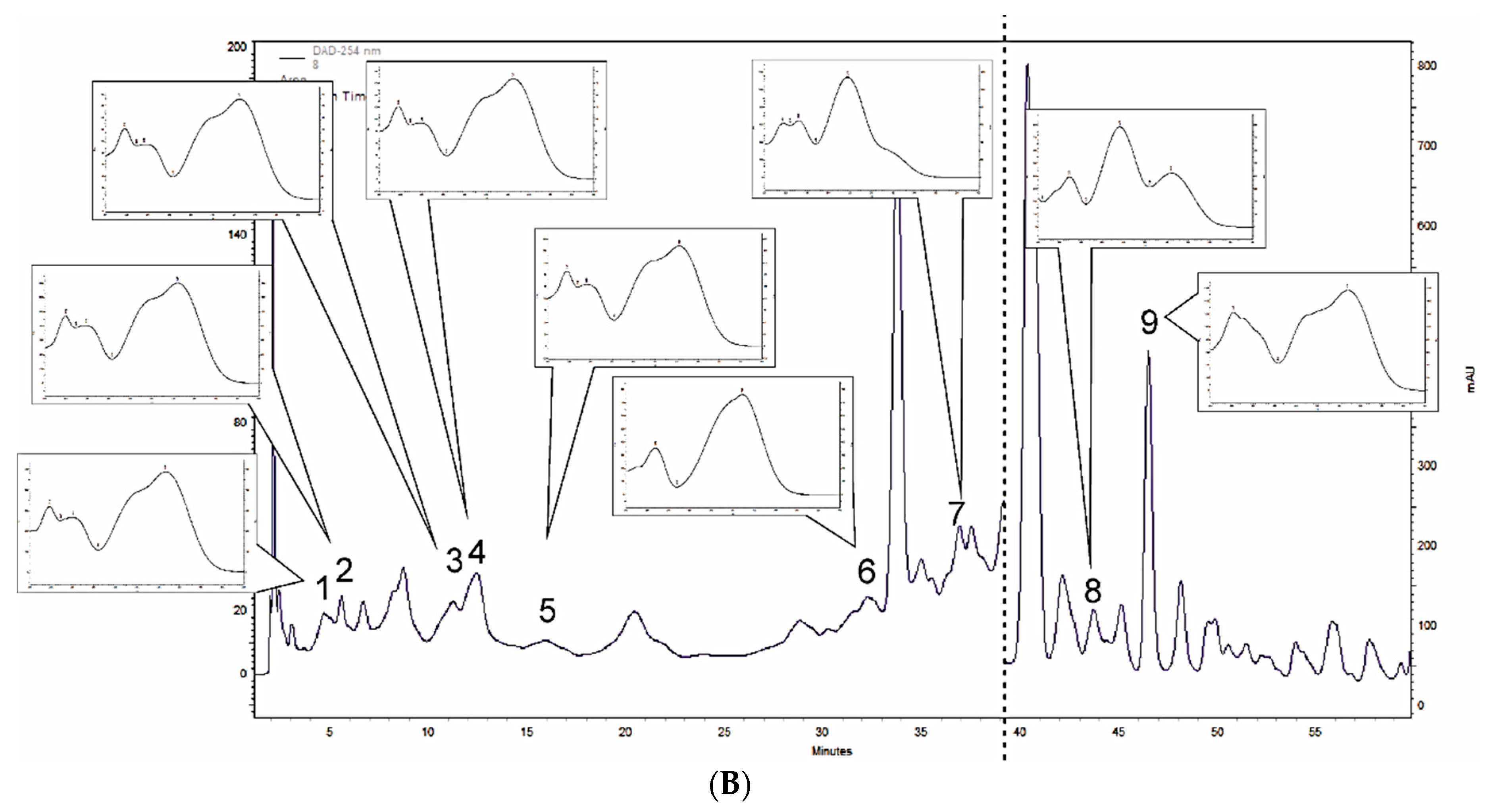
3.1. Polyphenol Profiling of M. × piperita and M. longifolia Leaf Extracts

The HPLC-DAD phenolic profile analyses revealed qualitative and quantitative differences in the M. × piperita and M. longifolia leaf extracts, as shown in Table 1 and Figure 1. In M. × piperita methanolic leaf extracts, six phenolic acids were detected (out of 26 screened compounds). Quantitatively, the major phenolic acid was rosmarinic acid (1547.6 mg/100 g DW dry weight (DW)), while the other phenolic acids were lower: cryptochlorogenic acid (91.7 mg/100 g DW), chlorogenic acid (69.4 mg /100 g DW). Protocatechuic acid, caffeic acid, and isochlorogenic acid were much lower. We screened for 16 flavonoids in the M. × piperita leaf extracts and three were confirmed: cynaroside (luteolin-7-glucoside) (162.8 mg/100 g DW) and naringin (naringenin 7-rhamnoglucoside) (328.8 mg/100 g DW), as shown in Table 1 and Figure 1A.
Nine phenolic acids were confirmed in the M. longifolia leaf extracts: rosmarinic acid (781.6 mg/100 g DW), cryptochlorogenic acid (191.1 mg/100 g DW), p-coumaric acid (113.0 mg/100 g DW), m-coumaric acid (112.2 mg/100 g DW), neochlorogenic acid (56.8 mg/100 g DW), caftaric acid (47.6 mg/100 g DW), chlorogenic acid (63.8 mg/100 g DW), o-coumaric acid (39.8 mg/100 g DW), and caffeic acid (19.6 mg/100 g DW), as displayed in Table 1 and Figure 1B. There were no confirmed flavonoids.

3.2. Antioxidant Effects

M. × piperita and M. longifolia leaf extracts and the individual compounds showed significant antioxidant activities, as presented in Table 2. M. longifolia showed higher antioxidant activities than M. × piperita using β-carotene bleaching assay. However, DPPH and FRAP assays did not show these differences at p ≤ 0.05. The phenolic compounds, rosmarinic acid, cynaroside, and cryptochlorogenic acid, had high antioxidant activities that were comparable to the BHT and Trolox standards. Naringin showed relatively low antioxidant activities compared to the other compounds and the leaf extracts.

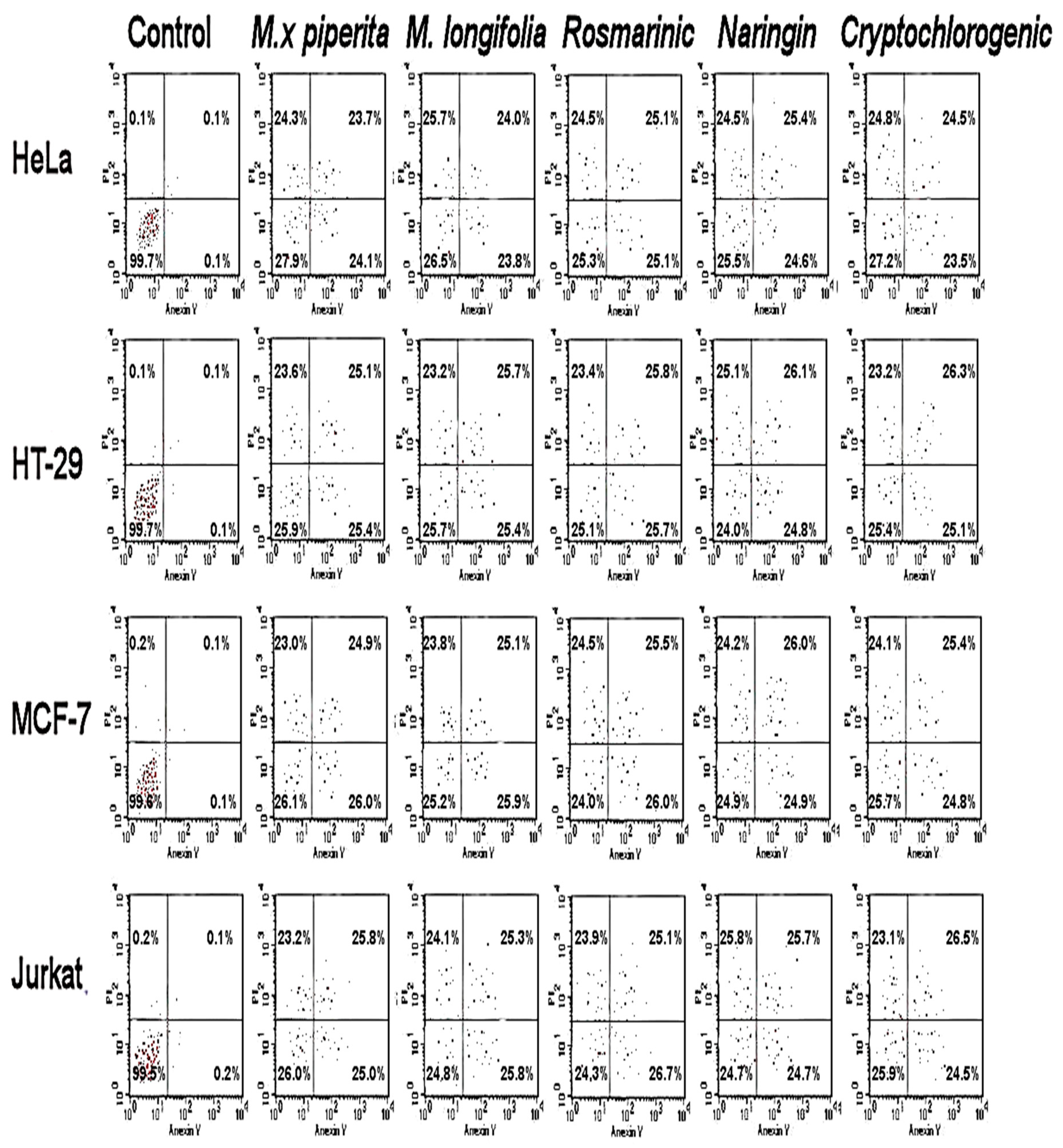
3.3. Antiproliferative and Cytotoxic Effects

Antiproliferative activities were measured against the Jurkat, MCF-7, HT-29, HEK-293, and HeLa cell lines. The leaf extracts of M. × piperita and M. longifolia showed antiproliferative activities against a diversity of cancer cells as shown in Table 3, and M. × piperita had higher activities than M. longifolia. Naringin, cryptochlorogenic acid, and rosmarinic acid had the highest activities among the identified compounds.
The apoptotic assay using flow cytometry of M. × piperita and M. longifolia is shown in Figure 2. Necrotic cell accumulation was observed in the early and late apoptotic periods. Necrotic and apoptotic cell accumulation was observed in the upper left (necrotic) and upper right and lower quadrants (apoptotic).

3.4. Antibacterial Activities

The leaf extracts of M. × piperita and M. longifolia showed variable antibacterial activities against the screened bacterial strains, as shown in Table 4. Most of the bacteria, including Pseudomonas aeruginosa, Micrococcus flavus, Bacillus cereus, Staphylococcus aureus, and Escherichia coli, were sensitive to both leaf extracts. Listeria monocytogenes was relatively sensitive to the leaf extracts, and M. × piperita showed higher antibacterial activity than M. longifolia. Among the individual compounds, caffeic and cryptochlorogenic acids had higher activities compared to rosmarinic acid and naringin.

3.5. Antifungal Effects

The leaf extracts of M. × piperita and M. longifolia showed antifungal effects against all of the screened bacteria including P. ochrochloron, P. funiculosum, A. ochraceus, A. niger, and A. flavus, as shown in Table 5. M. × piperita had higher activity than M. longifolia, and caffeic acid and naringin had the highest activities among the polyphenols. Cryptochlorogenic acid showed moderate effects against the fungi, while rosmarinic acid had the weakest antifungal activity. A. niger, A. ochraceus, and C. albicans were most resistant to the leaf extracts.

4. Discussion

Most phytochemical studies of Mentha have focused on analyses of the essential oil. Mentha’s volatile constituents are well-recognized; the main chemical constituent of the essential oil is menthol (33–60%) [40,54]. The other compounds are menthone (15–32%), 1,8-cineole (eucalyptol) (5–13%), menthofuran (1–10%), isomenthone (1.5–10.0%), menthyl acetate (2–11.0%), limonene (1–7%), and β-caryophyllene (2–4%) [26,30,54,55,56]. Prior work has suggested that Mentha’s essential oil is mainly responsible for the biological activity of the plant products [57,58]. Olennikov et al. [42] reported that the total phenolic compound contents of different Mentha species ranged from approximately 6% to 12%. This indicates that the phenolic compounds such as phenolic acids, flavonoids, and tannins play a significant role in the biological activities of the Mentha extracts [41]. The phenolic composition of Mentha’s leaves lacks complete description and even fewer studies have examined M. × piperita [40,41,59].
In this study, an HPLC–DAD phenolic profile analyses revealed the qualitative and quantitative differences of M. × piperita and M. longifolia methanolic leaf extracts, as shown in Table 1 and Figure 1. In M. × piperita extracts, six phenolic acids were detected, while in M. longifolia, nine were identified. Four depsides (cryptochlorogenic acid, chlorogenic acid, isochlorogenic acid, and rosmarinic acid) and two simple phenolic acids (caffeic acid and protocatechuic acid) were observed among the M. × piperita polyphenols. Among the M. longifolia polyphenols, there were five depsides (caftaric acid, chlorogenic acid, cryptochlorogenic acid, neochlorogenic acid, and rosmarinic acid) and four simple phenolic acids (caffeic acid, p-, m-, and o-coumaric acids). In both Mentha leaf extracts, rosmarinic acid was the major compound; at 1547.6 mg/100 g DW, it was almost two times higher in M. × piperita than in M. longifolia (781.6 mg/100 g DW). Earlier studies also reported rosmarinic acid as the main phenolic acid in Mentha species [60,61]. For M. × piperita specifically, caffeic acid and protocatechuic acid were also reported [40]. We confirmed the presence of these simple phenolic acids in the leaf extracts of M. × piperita, observing 17.4 and 24.9 mg/100 g DW, respectively. We also, for the first time, confirmed the presence of other biologically active depsides: cryptochlorogenic acid (91.7 mg/100 g DW) and isochlorogenic acid (10.3 mg/100 g DW). Among depsides, only chlorogenic acid was confirmed before in M. × piperita leaf extracts by Kapp et al. [62], but without quantification. In the current study we calculated the amount of this compound as 69.4 mg/100 g DW. Zgórka and Głowniak [63] performed the quantitative analyses of extracts from different plants belonging to the Lamiaceae family by HPLC with UV–Vis detection. They found rosmarinic acid (ca. 70 mg/100 g DW), caffeic acid (ca. 20 mg/100 g DW), and also protocatechuic acid (2.5 mg/100 g DW) in M. × piperita leaf extracts of Polish origin, as the dominant compounds. The amounts of rosmarinic acid and protocatechuic acid were respectively 22 and 10 times lower in comparison to our Saudi-origin samples. The amounts of caffeic acid were similar. The quantitative estimations of phenolic acid content in Serbian origin M. × piperita leaf extracts were performed by Mišan et al. [64], and they detected a 10 times higher amount of caffeic acid (186 mg/100 g DW), the similar amount of chlorogenic acid (73 mg/100 g DW) and an extremely lower—130 times—amount of rosmarinic acid (12 mg/100 g DW), in comparison to our study.
Inoue et al. [65] isolated flavonoids from the aerial sections of M. × piperita, including eriocitrin, luteolin-7-O-rutinoside, narirutin, hesperidin, isorhoifolin, diosmin, and 5,7-dihydroxycromone-7-O-rutinoside. There are also other flavonoids—chrysoeriol, luteolin, luteolin glucoside, naringenin glucoside, rutoside, nodifloretin, and eriodicytol, the most prominent of which are eriocitrin, luteolin 7-O-rutinoside, and hesperidin [40,41,66]. We screened for 16 flavonoid compounds using commercially available standards (see Materials and Methods) and observed two compounds in M. × piperita: cynaroside (luteolin-7-glucoside) (162.8 mg/100 g DW) and naringin (naringenin 7-rhamnoglucoside) (328.8 mg/100 g DW). These compounds are recognized for their structural similarity to luteolin-7-O-rutinoside (PubChem database: https://pubchem.ncbi.nlm.nih.gov/#query=CID14032966%20structure&tab=similarity).
Cynaroside (luteolin-7-glucoside) was confirmed in M. × piperita leaf extracts of Polish origin by Fecka et al. [67] in the amount of ca. 37 mg/100 g DW, using planar chromatography. That amount was 4.4 times lower in comparison to our samples of Saudi-origin plants. The naringin was detected by Figueroa Pérez et al. [68] in Mexican-origin M. × piperita leaf extracts at 56 mg/100 g DW, which was almost six times lower than detected under our study. Additionally, derivatives of naringin in M. × piperita were detected before by Mišan et al. [64] using the HPLC–DAD method. They confirmed naringenin in amount of 146 mg/100 g DW in the Serbian M. × piperita leaf extracts.
Phytochemically, M. longifolia is the less-known and less-studied species. Gulluce et al. [69] studied the essential oil and methanol extracts of M. longifolia ssp. longifolia. This resulted in the identification of 45 essential oil compounds; the main components were cis-piperitone epoxide, pulegone, piperitenone oxide, and menthone. The phenolic compound analyses were limited to spectrophotometric total phenolic constituent estimations using the Folin–Ciocalteu reagent, but the assay revealed total phenolic contents of 4.5 g/100 g of gallic acid equivalent. Akroum et al. [70] studied the flavonoid composition of M. longifolia extracts and identified five flavonoids (luteolin-7-O-glycoside, luteolin-7,3’-O-diglycoside, apigenin, quercetin-3-O-glycoside, and kaempferol-3-O-glycoside). We did not detect any flavonoids in M. longifolia, but we screened for only 16 flavonoids and used simple HPLC–DAD methods (see Materials and Methods). It is still possible that flavonoids are present in M. longifolia of Saudi origin, but in small amounts. In this study, we identified nine phenolic acids, including five depsides: rosmarinic acid (781.6 mg/100 g DW), cryptochlorogenic acid (191.1 mg/100 g DW), neochlorogenic acid (56.8 mg/100 g DW), chlorogenic acid (63.8 mg/100 g DW), and caftaric acid (47.6 mg/100 g DW), and four simple phenolic acids: caffeic acid (19.6 mg/100 g DW), p-coumaric acid (113.0 mg/100 g DW), m-coumaric acid (112.2 mg/100 g DW), and o-coumaric acid (39.8 mg/100 g DW). Adham [71], reported rosmarinic acid in an amount 3.75 times lower than in our samples (208 mg/100 g DW) in M. longifolia. The high amount of rosmarinic acid was also reported by Bahadori et al. in the M. longifolia var. calliantha [72] leaf extracts of plants growing in West Azerbaijan province of Iran. The amount of this depside was equal to 2225 µg/g in the ethanol extract. They confirmed also the presence of o-coumaric acid (134 µg/g), p-coumaric acid (113 µg/g), caffeic acid (86 µg/g) and chlorogenic acid (27 µg/g). Shekarchi et al. [73] with the HPLC–UV–Vis technique detected the rosmarinic acid in the leaf extracts of M. longifolia of Iranian origin in an amount of 2260 mg/100 g DW; that was 3.4 times more than in our Saudi plants. Dudai et al. [74] studied 40 genetic variants from 25 populations of M. longifolia native to Israel. They stated that rosmarinic and caffeic acids were the most abundant. The accumulation of rosmarinic acid, varied from 2000 to 8000 mg/100 g DW, caffeic acid from 60 to 1800 mg/100 g DW, and p-coumaric acid from 2.5 to 25 mg/100 g DW. These compounds were detected also in our study. The amounts of rosmarinic and caffeic acids were lower, but the amount of p-coumaric acid in the Saudi origin samples was higher.
We confirmed the availability of some polyphenols such as m-coumaric acid, neochlorogenic acid and caftaric acid in M. longifolia and cryptochlorogenic acid and isochlorogenic acid in M. × piperita in the Saudi origin plants for the first time (see Table S1 in the Supplementary Material). However, other phenols were reported in previous studies. Indeed, further investigations may reveal other compounds not confirmed here which could be considered as limitation of this study.
The DPPH, β-carotene bleaching, and FRAP assays showed higher antioxidant activities in M. longifolia than in M. × piperita. The phenolic compounds (rosmarinic acid, cynaroside, and cryptochlorogenic acid) also showed high antioxidant activities, while naringin showed relatively low activities compared to other compounds and leaf extracts. Our results for the antioxidant activity of rosmarinic acid via DPPH is consistent with prior work on Melissa officinalis [75]. Citrus junos extracts rich in naringin showed moderate antioxidant activities [76], but a different study documented weaker activities [77]. Cynaroside obtained from Elsholtzia bodinieri had strong antioxidant activities (via the DPPH assay) [78], and cryptochlorogenic and chlorogenic acids from Hibiscus sabdariffa showed strong antioxidant activities [79]. These results agree with the observations of our study.
M. × piperita showed higher activities against different cancer cells than M. longifolia, and the highest activities of the phenolic compounds were found in naringin, cryptochlorogenic acid, and rosmarinic acid. Previous work documented antiproliferative effects of rosemary leaf extracts against HT-29 cancer cells by inhibiting cell proliferation, increasing cell cycle arrest, and increasing apoptosis [80]. The major component of this extract was rosmarinic acid [81]. Rosmarinic acid has strong anticancer activities against MCF-7 [82] and HT-29 [83]. Naringin is a bioflavonoid that inhibits the growth of different cancer cells (e.g., MCF-7 [84]) and may induce apoptosis by reducing the NF-κB/COX-2-caspase-1 pathway in HeLa cells [85]. This explains the strong antiproliferative and cytotoxic activities reported here.
M. × piperita showed higher antibacterial activity than M. longifolia. Caffeic and cryptochlorogenic acids showed higher activities against bacteria compared to rosmarinic acid and naringin. M. × piperita also showed higher antifungal activity than M. longifolia, and caffeic acid and naringin had the highest antifungal activities among the polyphenols. Cryptochlorogenic acid was moderately effective against fungi, but rosmarinic acid was the weakest. Previous studies reported moderate-to-low antimicrobial activities for rosmarinic acid [86,87] and naringin [88], and high antibacterial activities have been reported for chlorogenic acid (cryptochlorogenic acid derivatives) from Mammillaria extracts [4,89]. Chlorogenic acid displayed fungicidal activities against other pathogenic fungi such as Fusarium solani, Sclerotinia sclerotiorum, and Botrytis cinerea [90], and caffeic acid showed strong antibacterial and antifungal activities against several bacterial strains [2].

5. Conclusions

This is the first investigation of the phenol profiles of natural Mentha populations from the Riyadh region in northern Saudi Arabia. HPLC identified several polyphenols including rosmarinic acid, naringin, cryptochlorogenic acid, and cymaroside. The main component of the polyphenols in M. × piperita and M. longifolia was rosmarinic acid. M. × piperita contained flavonoids (naringin and cymaroside), and other polyphenols were present in M. longifolia, including cryptochlorogenic, m-coumaric acid, and p-coumaric acid. Most of the phenols showed antioxidant activities and were associated with the antioxidant activities of the leaf extracts. The M. × piperita and M. longifolia leaf extracts displayed antiproliferative and cytotoxic effects against most of the cancer cell lines, which was attributed to the accumulation of necrotic cells in the early and late apoptotic periods. Antibacterial effects were detected in both of the leaf extracts and in the polyphenols and were effective against most of the bacteria. Furthermore, M. × piperita showed higher antibacterial activity than M. longifolia. This activity was attributed to the specific polyphenol profile of the plant (i.e., cryptochlorogenic and caffeic acids and twice the amount of rosmarinic acid). Antifungal activities were also detected for both species and were attributed to the polyphenols caffeic acid, cryptochlorogenic acid, and naringin. This is the first study confirming the availability of some polyphenols such as m-coumaric acid, neochlorogenic acid, caftaric acid in M. Longifolia and cryptochlorogenic acid and isochlorogenic acid in M. x Piperita. Together, these results indicate that both mint species may represent new natural sources of biologically active polyphenols.

Supplementary Materials

The following are available online at https://0-www-mdpi-com.brum.beds.ac.uk/2227-9717/8/4/479/s1, Table S1: The comparison of detected polyphenolic compounds under our study with the results of other teams.

Author Contributions

Conceptualization, H.O.E., A.S., D.O.E.-A. and E.A.M.; data curation, H.O.E., A.S., P.K., M.K.-S, D.O.E.-A. and E.A.M.; formal analysis, H.O.E., H.E., P.K., H.E., M.K.-S., D.O.E.-A. and E.A.M.; funding acquisition, H.O.E., A.S., H.E., and D.O.E.-A.; investigation, H.O.E., A.S., P.K., M.K.-S., D.O.E.-A. and E.A.M.; methodology, H.O.E., A.S., P.K. and M.K.-S.; visualization, H.O.E., A.S., P.K.; writing—original draft, H.O.E., and A.S.; writing—review and editing, H.O.E. and A.S. All authors have read and agreed to the published version of the manuscript.

Funding

This work was supported by King Saud University, Researchers Supporting Project number: RSP-2019/118.

Acknowledgments

The authors extend their appreciation to King Saud University, Researchers Supporting Project (RSP-2019/118) for funding this work.

Conflicts of Interest

The authors declare no conflict of interest.

References

  1. Elansary, H.O.; Szopa, A.; Kubica, P.; Al-Mana, F.A.; Mahmoud, E.A.; El-Abedin, T.Z.; Mattar, M.A.; Ekiert, H.; El-Abedin, T.K.A.Z. Phenolic Compounds of Catalpa speciosa, Taxus cuspidata, and Magnolia acuminata have Antioxidant and Anticancer Activity. Molecules 2019, 24, 412. [Google Scholar] [CrossRef] [Green Version]
  2. Elansary, H.O.; Szopa, A.; Kubica, P.; Ekiert, H.; Mattar, M.A.; Al-Yafrasi, M.A.; El-Ansary, D.O.; Zin El-Abedin, T.K.; Yessoufou, K. Polyphenol Profile and Pharmaceutical Potential of Quercus spp. Bark Extracts. Plants 2019, 8, 486. [Google Scholar] [CrossRef] [PubMed] [Green Version]
  3. Elansary, H.O. Tree Bark Phenols Regulate the Physiological and Biochemical Performance of Gladiolus Flowers. Processes 2020, 8, 71. [Google Scholar] [CrossRef] [Green Version]
  4. Elansary, H.O.; Szopa, A.; Klimek-Szczykutowicz, M.; Jafernik, K.; Ekiert, H.; Mahmoud, E.; Barakat, A.A.; El-Ansary, D.O. Mammillaria Species—Polyphenols Studies and Anti-Cancer, Anti-Oxidant, and Anti-Bacterial Activities. Molecules 2019, 25, 131. [Google Scholar] [CrossRef] [Green Version]
  5. Okla, M.; Alamri, S.; Salem, M.Z.M.; Ali, H.M.; Behiry, S.I.; Nasser, R.A.; Alaraidh, I.A.; Al-Ghtani, S.M.; Soufan, W. Yield, Phytochemical Constituents, and Antibacterial Activity of Essential Oils from the Leaves/Twigs, Branches, Branch Wood, and Branch Bark of Sour Orange (Citrus aurantium L.). Processes 2019, 7, 363. [Google Scholar] [CrossRef] [Green Version]
  6. Salem, M.Z.M.; Elansary, H.O.; Ali, H.M.; El-Settawy, A.A.; Elshikh, M.S.; Abdel-Salam, E.M.; Skalicka-Wozniak, K. Bioactivity of essential oils extracted from Cupressus macrocarpa branchlets and Corymbia citriodora leaves grown in Egypt. BMC Complement. Altern. Med. 2018, 18, 23. [Google Scholar] [CrossRef]
  7. Halagarda, M.; Groth, S.; Popek, S.; Rohn, S.; Pedan, V. Antioxidant Activity and Phenolic Profile of Selected Organic and Conventional Honeys from Poland. Antioxidants 2020, 9, 44. [Google Scholar] [CrossRef] [Green Version]
  8. Wang, S.; Moustaid-Moussa, N.; Chen, L.; Mo, H.; Shastri, A.; Su, R.; Bapat, P.; Kwun, I.; Shen, C.-L. Novel insights of dietary polyphenols and obesity. J. Nutr. Biochem. 2014, 25, 1–18. [Google Scholar] [CrossRef] [Green Version]
  9. Ebrahimi, A.; Schluesener, H. Natural polyphenols against neurodegenerative disorders: Potentials and pitfalls. Ageing Res. Rev. 2012, 11, 329–345. [Google Scholar] [CrossRef]
  10. Cao, H.; Ou, J.; Chen, L.; Zhang, Y.; Szkudelski, T.; Delmas, M.; Daglia, M.; Ulrih, N.P. Dietary polyphenols and type 2 diabetes: Human Study and Clinical Trial. Crit. Rev. Food Sci. Nutr. 2018, 59, 3371–3379. [Google Scholar] [CrossRef]
  11. Tangney, C.; Rasmussen, H. Polyphenols, inflammation, and cardiovascular disease. Curr. Atheroscler. Rep. 2013, 15, 324. [Google Scholar] [CrossRef] [PubMed]
  12. Magrone, T.; Magrone, M.; Russo, M.A.; Jirillo, E. Magrone Recent Advances on the Anti-Inflammatory and Antioxidant Properties of Red Grape Polyphenols: In Vitro and In Vivo Studies. Antioxidants 2019, 9, 35. [Google Scholar] [CrossRef] [PubMed] [Green Version]
  13. Khan, M.; Siddiqui, S. Concurrent chemoradiotherapy with or without induction chemotherapy for the management of cervical lymph node metastasis from unknown primary tumor. J. Cancer Res. Ther. 2018, 14, 1117. [Google Scholar] [CrossRef]
  14. Alvarado-Sansininea, J.J.; Sánchez, L.S.; López-Muñoz, H.; Escobar, M.L.; Flores-Guzmán, F.; Tavera-Hernández, R.; Jimenez-Estrada, M. Quercetagetin and Patuletin: Antiproliferative, Necrotic and Apoptotic Activity in Tumor Cell Lines. Molecules 2018, 23, 2579. [Google Scholar] [CrossRef] [Green Version]
  15. Sezer, E.D.; Oktay, L.M.; Karadadaş, E.; Memmedov, H.; Gunel, N.S.; Sözmen, E. Assessing Anticancer Potential of Blueberry Flavonoids, Quercetin, Kaempferol, and Gentisic Acid, Through Oxidative Stress and Apoptosis Parameters on HCT-116 Cells. J. Med. Food 2019, 22, 1118–1126. [Google Scholar] [CrossRef]
  16. Yessoufou, K.; Daru, B.H.; Tafirei, R.; Elansary, O.H.; Rampedi, I. Integrating biogeography, threat and evolutionary data to explore extinction crisis in the taxonomic group of cycad. Ecol. Evol. 2017, 7, 2735–2746. [Google Scholar] [CrossRef] [Green Version]
  17. Saleh, A.; Elfayoumi, H.M.; Youns, M.; Barakat, W. Rutin and orlistat produce antitumor effects via antioxidant and apoptotic actions. Naunyn-Schmiedeberg’s Arch. Pharmacol. 2018, 392, 165–175. [Google Scholar] [CrossRef]
  18. Dayem, A.A.; Choi, H.Y.; Yang, G.-M.; Kim, K.; Saha, S.K.; Cho, S.G. The Anti-Cancer Effect of Polyphenols against Breast Cancer and Cancer Stem Cells: Molecular Mechanisms. Nutrients 2016, 8, 581. [Google Scholar] [CrossRef]
  19. Costea, T.; Nagy, P.; Ganea, C.; Szöllősi, J.; Mocanu, M.-M. Molecular Mechanisms and Bioavailability of Polyphenols in Prostate Cancer. Int. J. Mol. Sci. 2019, 20, 1062. [Google Scholar] [CrossRef] [Green Version]
  20. Kim, H.K.; Missiakas, M.; Schneewind, O. Mouse models for infectious diseases caused by Staphylococcus aureus. J. Immunol. Methods 2014, 410, 88–99. [Google Scholar] [CrossRef]
  21. Mahmoud, E.A.; Elansary, H.O.; Al-Mana, F.A.; Al-Mana, F.A. Elevated Bioactivity of Ruta graveolens against Cancer Cells and Microbes Using Seaweeds. Processes 2020, 8, 75. [Google Scholar] [CrossRef] [Green Version]
  22. Desai, A.N.; Anyoha, A.; Madoff, L.C.; Lassmann, B. Changing epidemiology of Listeria monocytogenes outbreaks, sporadic cases, and recalls globally: A review of ProMED reports from 1996 to 2018. Int. J. Infect. Dis. 2019, 84, 48–53. [Google Scholar] [CrossRef] [PubMed] [Green Version]
  23. Elansary, H.O.; Szopa, A.; Kubica, P.; Ekiert, H.; Ali, H.M.; Elshikh, M.S.; Abdel-Salam, E.M.; El-Esawi, M.A.; El-Ansary, D.O. Bioactivities of Traditional Medicinal Plants in Alexandria. Evid.-Based Complement. Altern. Med. 2018, 2018, 1–13. [Google Scholar] [CrossRef] [PubMed] [Green Version]
  24. Elansary, H.O.; Salem, M.Z.M.; Ashmawy, N.A.; Yessoufou, K.; El-Settawy, A.A. In vitro antibacterial, antifungal and antioxidant activities of Eucalyptus spp. leaf extracts related to phenolic composition. Nat. Prod. Res. 2017, 9, 1–4. [Google Scholar] [CrossRef] [PubMed]
  25. Ventola, C.L. The antibiotic resistance crisis: Part 1: Causes and threats. Pharm. Ther. 2015, 40, 277–283. [Google Scholar]
  26. Elansary, H.O.; El-Abedin, T.K.Z. Omeprazole alleviates water stress in peppermint and modulates the expression of menthol biosynthesis genes. Plant Physiol. Biochem. 2019, 139, 578–586. [Google Scholar] [CrossRef]
  27. Elansary, H.O.; Mahmoud, E.A.; El-Ansary, D.O.; Mattar, M.A. Effects of Water Stress and Modern Biostimulants on Growth and Quality Characteristics of Mint. Agronomy 2019, 10, 6. [Google Scholar] [CrossRef] [Green Version]
  28. Elansary, H.O. Green roof Petunia, Ageratum, and Mentha responses to water stress, seaweeds, and trinexapac-ethyl treatments. Acta Physiol. Plant. 2017, 39, 145. [Google Scholar] [CrossRef]
  29. Elansary, H.O.; Yessoufou, K.; Shokralla, S.; Mahmoud, E.; Skalicka-Wozniak, K. Enhancing mint and basil oil composition and antibacterial activity using seaweed extracts. Ind. Crop. Prod. 2016, 92, 50–56. [Google Scholar] [CrossRef]
  30. Elansary, H.O.; Ashmawy, N.A. Essential Oils of Mint between Benefits and Hazards. J. Essent. Oil Bear. Plants 2013, 16, 429–438. [Google Scholar] [CrossRef]
  31. McKay, D.L.; Blumberg, J.B. A review of the bioactivity and potential health benefits of peppermint tea (Mentha piperita L.). Phytother. Res. 2006, 20, 619–633. [Google Scholar] [CrossRef] [PubMed]
  32. Xhang, X. WHO Monographs on Selected Medicinal Plants WHO: Essential Medicines and Health Products Information Portal; World Health Organization: Geneva, Switzerland, 2004; Volume 2, p. 358. [Google Scholar]
  33. Medicines, E.D.f.t.Q.o. European Pharmacopoeia 9.0; Council of Europe: Strasburg, Germany, 2017. [Google Scholar]
  34. Mkaddem, M.; Bouajila, J.; Ennajar, M.; Lebrihi, A.; Mathieu, F.; Romdhane, M. Chemical Composition and Antimicrobial and Antioxidant Activities of Mentha (longifolia L. and viridis) Essential Oils. J. Food Sci. 2009, 74, M358–M363. [Google Scholar] [CrossRef] [PubMed]
  35. Moloudizargari, M.; Aghajanshakeri, S.; Mikaili, P.; Mojaverrostami, S. Pharmacological and therapeutic effects of Mentha longifolia L. and its main constituent, menthol. Anc. Sci. Life 2013, 33, 129–138. [Google Scholar] [CrossRef] [PubMed] [Green Version]
  36. Zade, N.S.E.; Sadeghi, A.; Moradi, P. Streptomyces strains alleviate water stress and increase peppermint (Mentha piperita) yield and essential oils. Plant Soil 2018, 434, 441–452. [Google Scholar] [CrossRef]
  37. Rahimi, Y.; Taleei, A.; Ranjbar, M. Long-term water deficit modulates antioxidant capacity of peppermint (Mentha piperita L.). Sci. Hortic. 2018, 237, 36–43. [Google Scholar] [CrossRef]
  38. Elansary, H.O.; Mahmoud, E. Egyptian herbal tea infusions’ antioxidants and their antiproliferative and cytotoxic activities against cancer cells. Nat. Prod. Res. 2014, 29, 474–479. [Google Scholar] [CrossRef]
  39. Kamatou, G.P.; Vermaak, I.; Viljoen, A.; Lawrence, B.M. Menthol: A simple monoterpene with remarkable biological properties. Phytochemistry 2013, 96, 15–25. [Google Scholar] [CrossRef]
  40. Baliga, M.; Rao, S. Radioprotective potential of mint: A brief review. J. Cancer Res. Ther. 2010, 6, 255. [Google Scholar] [CrossRef]
  41. Fecka, I.; Kowalczyk, A.; Cisowski, W. Optimization of the separation of flavonoid glycosides and rosmarinic acid from Mentha piperita on HPTLC plates. J. Planar Chromatogr. Mod. TLC 2004, 17, 22–25. [Google Scholar] [CrossRef]
  42. Olennikov, D.N.; Tankhaeva, L.M. Quantitative determination of phenolic compounds in Mentha piperita leaves. Chem. Nat. Compd. 2010, 46, 22–27. [Google Scholar] [CrossRef]
  43. Elansary, H.O.M.; Adamec, L.; Štorchová, H. Uniformity of organellar DNA in Aldrovanda vesiculosa, an endangered aquatic carnivorous species, distributed across four continents. Aquat. Bot. 2010, 24, 214–220. [Google Scholar] [CrossRef]
  44. Sułkowska-Ziaja, K.; Maślanka, A.; Szewczyk, A.; Muszyńska, B. Physiologically Active Compounds in Four Species of Phellinus. Nat. Prod. Commun. 2017, 12, 363–366. [Google Scholar] [CrossRef] [PubMed] [Green Version]
  45. Szopa, A.; Kokotkiewicz, A.; Bednarz, M.; Luczkiewicz, M.; Ekiert, H. Studies on the accumulation of phenolic acids and flavonoids in different in vitro culture systems of Schisandra chinensis (Turcz.) Baill. using a DAD-HPLC method. Phytochem. Lett. 2017, 20, 462–469. [Google Scholar] [CrossRef]
  46. Yessoufou, K.; Elansary, H.O.; Mahmoud, E.; Skalicka-Wozniak, K. Antifungal, antibacterial and anticancer activities of Ficus drupacea L. stem bark extract and biologically active isolated compounds. Ind. Crop. Prod. 2015, 74, 752–758. [Google Scholar] [CrossRef]
  47. Elansary, H.O.; Abdelgaleil, S.A.; Mahmoud, E.; Yessoufou, K.; Elhindi, K.; El-Hendawy, S. Effective antioxidant, antimicrobial and anticancer activities of essential oils of horticultural aromatic crops in northern Egypt. BMC Complement. Altern. Med. 2018, 18, 214. [Google Scholar] [CrossRef]
  48. Elansary, H.O.; Yessoufou, K.; Abdel-Hamid, A.; El-Esawi, M.A.; Ali, H.M.; Elshikh, M.S. Seaweed Extracts Enhance Salam Turfgrass Performance during Prolonged Irrigation Intervals and Saline Shock. Front. Plant Sci. 2017, 8, 830. [Google Scholar] [CrossRef] [Green Version]
  49. Ferreira, J.; Miranda, I.; Sousa, V.; Pereira, H. Chemical composition of barks from Quercus faginea trees and characterization of their lipophilic and polar extracts. PLoS ONE 2018, 13, e0197135. [Google Scholar] [CrossRef]
  50. El-Esawi, M.A.; Elkelish, A.; Soliman, M.H.; Elansary, H.O.; Zaid, A.; Wani, S.H. Serratia marcescens BM1 Enhances Cadmium Stress Tolerance and Phytoremediation Potential of Soybean Through Modulation of Osmolytes, Leaf Gas Exchange, Antioxidant Machinery, and Stress-Responsive Genes Expression. Antioxidants 2020, 9, 43. [Google Scholar] [CrossRef] [Green Version]
  51. Elansary, H.O.; Szopa, A.; Klimek-Szczykutowicz, M.; Ekiert, H.; Barakat, A.A.; Al-Mana, F.A. Antiproliferative, Antimicrobial, and Antifungal Activities of Polyphenol Extracts from Ferocactus Species. Processes 2020, 8, 138. [Google Scholar] [CrossRef] [Green Version]
  52. Elansary, H.O.; Mahmoud, E. Basil cultivar identification using chemotyping still favored over genotyping using core barcodes and possible resources of antioxidants. J. Essent. Oil Res. 2014, 27, 82–87. [Google Scholar] [CrossRef]
  53. El-Kareem, M.S.A.; Rabbih, M.A.; Elansary, H.O.; Al-Mana, F.A. Mass Spectral Fragmentation of Pelargonium graveolens Essential Oil Using GC–MS Semi-Empirical Calculations and Biological Potential. Processes 2020, 8, 128. [Google Scholar] [CrossRef] [Green Version]
  54. Chumpitazi, B.P.; Kearns, G.L.; Shulman, R.J. Review article: The physiological effects and safety of peppermint oil and its efficacy in irritable bowel syndrome and other functional disorders. Aliment. Pharmacol. Ther. 2018, 47, 738–752. [Google Scholar] [CrossRef] [PubMed] [Green Version]
  55. Aziz, M.; Sharma, S.; Ghazaleh, S.; Fatima, R.; Acharya, A.; Ghanim, M.; Sheikh, T.; Lee-Smith, W.; Hamdani, S.U.; Nawras, A. The anti-spasmodic effect of peppermint oil during colonoscopy: A systematic review and meta-analysis. Minerva Gastroenterol. Dietol. 2020. [Google Scholar] [CrossRef]
  56. Keifer, D.; Ulbricht, C.; Abrams, T.R.; Basch, E.; Giese, N.; Giles, M.; Kirkwood, C.D.; Miranda, M.; Woods, J. Peppermint (Mentha X piperita). J. Herb. Pharmacother. 2008, 7, 91–143. [Google Scholar] [CrossRef]
  57. Salem, M.Z.M.; El-Hefny, M.; Nasser, R.A.; Ali, H.M.; El-Shanhorey, N.A.; Elansary, H.O. Medicinal and biological values of Callistemon viminalis extracts: History, current situation and prospects. Asian Pac. J. Trop. Med. 2017, 10, 229–237. [Google Scholar] [CrossRef] [PubMed]
  58. Badea, M.L.; Iconaru, S.; Groza, A.; Chifiriuc, M.C.; Beuran, M.; Predoi, M. Peppermint Essential Oil-Doped Hydroxyapatite Nanoparticles with Antimicrobial Properties. Molecules 2019, 24, 2169. [Google Scholar] [CrossRef] [PubMed] [Green Version]
  59. Mahendran, G.; Rahman, L. Ethnomedicinal, phytochemical and pharmacological updates on Peppermint (Mentha × piperita L.)-A review. Phytother. Res. 2020. [Google Scholar] [CrossRef] [PubMed]
  60. Ellis, B.E.; Towers, G.H.N. Biogenesis of rosmarinic acid in Mentha. Biochem. J. 1970, 118, 291–297. [Google Scholar] [CrossRef] [Green Version]
  61. Guedon, D.J.; Pasquier, B.P. Analysis and Distribution of Flavonoid Glycosides and Rosmarinic Acid in 40 Mentha x piperita Clones. J. Agric. Food Chem. 1994, 42, 679–684. [Google Scholar] [CrossRef]
  62. Kapp, K.; Hakala, E.; Orav, A.; Pohjala, L.; Vuorela, P.; Püssa, T.; Vuorela, H.; Raal, A. Commercial peppermint (Mentha × piperita L.) teas: Antichlamydial effect and polyphenolic composition. Food Res. Int. 2013, 53, 758–766. [Google Scholar] [CrossRef]
  63. Zgórka, G.; Głowniak, K. Variation of free phenolic acids in medicinal plants belonging to the Lamiaceae family. J. Pharm. Biomed. Anal. 2001, 26, 79–87. [Google Scholar] [CrossRef]
  64. Mišan, A.; Mimica-Dukić, N.M.; Mandić, A.I.; Sakač, M.B.; Milovanović, I.L.; Sedej, I.J. Development of a rapid resolution HPLC method for the separation and determination of 17 phenolic compounds in crude plant extracts. Open Chem. 2011, 9, 133–142. [Google Scholar] [CrossRef]
  65. Inoue, T.; Sugimoto, Y.; Masuda, H.; Kamei, C. Antiallergic Effect of Flavonoid Glycosides Obtained from Mentha piperita L. Boil. Pharm. Bull. 2002, 25, 256–259. [Google Scholar] [CrossRef] [PubMed] [Green Version]
  66. Kosar, M.; Dorman, H.J.D.; Baser, K.H.C.; Hiltunen, R. Screening of Free Radical Scavenging Compounds in Water Extracts of Mentha Samples Using a Postcolumn Derivatization Method. J. Agric. Food Chem. 2004, 52, 5004–5010. [Google Scholar] [CrossRef]
  67. Fecka, I.; Raj, D.; Krauze-Baranowska, M. Quantitative Determination of Four Water-Soluble Compounds in Herbal Drugs from Lamiaceae Using Different Chromatographic Techniques. Chromatographia 2007, 66, 87–93. [Google Scholar] [CrossRef]
  68. Pérez, M.G.F.; Rocha-Guzmán, N.E.; Mercado-Silva, E.; Loarca-Piña, G.; Reynoso–Camacho, R. Effect of chemical elicitors on peppermint (Mentha piperita) plants and their impact on the metabolite profile and antioxidant capacity of resulting infusions. Food Chem. 2014, 156, 273–278. [Google Scholar] [CrossRef]
  69. Gulluce, M.; Sahin, F.; Sokmen, M.; Ozer, H.; Daferera, D.; Sökmen, A.; Polissiou, M.; Adiguzel, A.; Ozkan, H. Antimicrobial and antioxidant properties of the essential oils and methanol extract from Mentha longifolia L. ssp. longifolia. Food Chem. 2007, 103, 1449–1456. [Google Scholar] [CrossRef]
  70. Akroum, S.; Bendjeddou, D.; Satta, D.; Lalaoui, K. Antibacterial activity and acute toxicity effect of flavonoids extracted from Mentha longifolia. Am.-Eurasian J. Sci. Res. 2009, 4, 93–96. [Google Scholar]
  71. Adham, A.N. Comparative extraction methods, phytochemical constituents, fluorescence analysis and HPLC validation of rosmarinic acid content in Mentha piperita, Mentha longifolia and Osimum basilicum. J. Pharmacogn. Phytochem. 2015, 3, 130–139. [Google Scholar]
  72. Zengin, G.; Bahadori, S.; Dinparast, L.; Movahhedin, N. Phenolic composition and functional properties of wild mint (Mentha longifolia var. calliantha (Stapf) Briq.). Int. J. Food Prop. 2018, 21, 183–193. [Google Scholar] [CrossRef] [Green Version]
  73. Shekarchi, M.; Hajimehdipoor, H.; Saeidnia, S.; Gohari, A.; Hamedani, M. Comparative study of rosmarinic acid content in some plants of Labiatae family. Pharmacogn. Mag. 2012, 8, 37–41. [Google Scholar] [CrossRef] [PubMed] [Green Version]
  74. Dudai, N.; Segey, D.; Haykin-Frenkel, D.; Eshel, A. Genetic Variation of Phenolic Compounds Content, Essential Oil Composition and Anti Oxidative Activity in Israel-Grown Mentha longifolia L. Acta Hortic. 2006, 69–78. [Google Scholar] [CrossRef]
  75. Canelas, V.; Teixeira da Costa, C. Quantitative HPLC Analysis of Rosmarinic Acid in Extracts of Melissa officinalis and Spectrophotometric Measurement of Their Antioxidant Activities. J. Chem. Educ. 2007, 84, 1502. [Google Scholar] [CrossRef]
  76. Shim, J.-H.; Chae, J.-I.; Cho, S.-S. Identification and Extraction Optimization of Active Constituents in Citrus junos Seib ex TANAKA Peel and Its Biological Evaluation. Molecules 2019, 24, 680. [Google Scholar] [CrossRef] [PubMed] [Green Version]
  77. De Martino, L.; Mencherini, T.; Mancini, E.; Aquino, R.P.; De Almeida, L.F.R.; De Feo, V. In Vitro Phytotoxicity and Antioxidant Activity of Selected Flavonoids. Int. J. Mol. Sci. 2012, 13, 5406–5419. [Google Scholar] [CrossRef] [PubMed]
  78. Zou, Y.; Zhang, M.; Zhang, T.; Wu, J.; Wang, J.; Liu, K.; Zhan, N. Antioxidant and Anti-inflammatory Activities of Cynaroside from Elsholtiza bodinieri. Nat. Prod. Commun. 2018, 13. [Google Scholar] [CrossRef] [Green Version]
  79. Wang, J.; Cao, X.; Jiang, H.; Qi, Y.; Chin, K.L.; Yue, Y.-D. Antioxidant Activity of Leaf Extracts from Different Hibiscus sabdariffa Accessions and Simultaneous Determination Five Major Antioxidant Compounds by LC-Q-TOF-MS. Molecules 2014, 19, 21226–21238. [Google Scholar] [CrossRef] [Green Version]
  80. Valdés, A.; Sullini, G.; Ibáñez, E.; Cifuentes, A.; Garcia-Cañas, V. Rosemary polyphenols induce unfolded protein response and changes in cholesterol metabolism in colon cancer cells. J. Funct. Foods 2015, 15, 429–439. [Google Scholar] [CrossRef]
  81. Afonso, M.; Silva, A.; Carvalho, E.B.; Rivelli, D.P.; Barros, S.B.D.M.; Rogero, M.M.; Lottenberg, A.; Torres, R.P.; Mancini-Filho, J. Phenolic compounds from Rosemary (Rosmarinus officinalis L.) attenuate oxidative stress and reduce blood cholesterol concentrations in diet-induced hypercholesterolemic rats. Nutr. Metab. 2013, 10, 19. [Google Scholar] [CrossRef] [Green Version]
  82. Berdowska, I.; Zielinski, B.; Fecka, I.; Kulbacka, J.; Saczko, J.; Gamian, A. Cytotoxic impact of phenolics from Lamiaceae species on human breast cancer cells. Food Chem. 2013, 141, 1313–1321. [Google Scholar] [CrossRef]
  83. Scheckel, K.A.; Degner, S.C.; Romagnolo, D.F. Rosmarinic acid antagonizes activator protein-1-dependent activation of cyclooxygenase-2 expression in human cancer and nonmalignant cell lines. J. Nutr. 2008, 138, 2098–2105. [Google Scholar] [CrossRef] [PubMed]
  84. Rajamani, S.; Radhakrishnan, A.; Sengodan, T.; Thangavelu, S. Augmented anticancer activity of naringenin-loaded TPGS polymeric nanosuspension for drug resistive MCF-7 human breast cancer cells. Drug Dev. Ind. Pharm. 2018, 44, 1752–1761. [Google Scholar] [CrossRef] [PubMed]
  85. Zeng, L.; Zhen, Y.; Chen, Y.; Zou, L.; Zhang, Y.; Hu, F.; Feng, J.; Shen, J.; Wei, B. Naringin inhibits growth and induces apoptosis by a mechanism dependent on reduced activation of NF-κB/COX-2-caspase-1 pathway in HeLa cervical cancer cells. Int. J. Oncol. 2014, 45, 1929–1936. [Google Scholar] [CrossRef] [PubMed] [Green Version]
  86. Matejczyk, M.; Świsłocka, R.; Golonko, A.; Lewandowski, W.; Hawrylik, E. Cytotoxic, genotoxic and antimicrobial activity of caffeic and rosmarinic acids and their lithium, sodium and potassium salts as potential anticancer compounds. Adv. Med Sci. 2018, 63, 14–21. [Google Scholar] [CrossRef] [PubMed]
  87. Fialová, S.; Kello, M.; Čoma, M.; Slobodníková, L.; Drobná, E.; Holková, I.; Garajová, M.; Garajová, M.; Zachar, V.; Lukáč, M. Derivatization of Rosmarinic Acid Enhances its in vitro Antitumor, Antimicrobial and Antiprotozoal Properties. Molecules 2019, 24, 1078. [Google Scholar] [CrossRef] [Green Version]
  88. Celiz, G.; Daz, M.; Audisio, M. Antibacterial activity of naringin derivatives against pathogenic strains. J. Appl. Microbiol. 2011, 111, 731–738. [Google Scholar] [CrossRef] [Green Version]
  89. Lou, Z.; Wang, H.; Zhu, S.; Ma, C.; Wang, Z. Antibacterial Activity and Mechanism of Action of Chlorogenic Acid. J. Food Sci. 2011, 76, M398–M403. [Google Scholar] [CrossRef]
  90. Martínez, G.; Regente, M.; Jacobi, S.; Del Rio, M.; Pinedo, M.; De La Canal, L.; De Rio, M. Chlorogenic acid is a fungicide active against phytopathogenic fungi. Pestic. Biochem. Physiol. 2017, 140, 30–35. [Google Scholar] [CrossRef]
Figure 1. Examples of high-performance liquid chromatography with diode-array detection (HPLC-DAD) chromatographic separation (λ = 254 nm) for the leaf extracts of (A) Mentha × piperita (1—protocatechuic acid; 2—cryptochlorogenic acid; 3—chlorogenic acid; 4—caffeic acid; 5—isochlorogenic acid; 6—cynaroside; 7—naringin; 8—rosmarinic acid) and (B) Mentha longifolia (1—caftaric acid; 2—neochlorogenic acid; 3—cryptochlorogenic acid; 4—chlorogenic acid; 5—caffeic acid; 6—p-coumaric acid; 7—m-coumaric acid; 8—o-coumaric acid; 9—rosmarinic acid).
Processes 08 00479 g001aProcesses 08 00479 g001b
Figure 2. Cytotoxicity of the M. × piperita and M. longifolia leaf extracts and naringin, rosmarinic acid, and cryptochlorogenic acid, estimated with flow cytometry. There was accumulation of apoptotic cells in the early (lower right quadrant) and late apoptotic (upper right quadrant).
Processes 08 00479 g002
Table 1. The quantitative estimates of polyphenols in M. × piperita and M. longifolia leaf extracts (mg/100 g DW dry weight (DW) ± standard deviation (SD)).
CompoundM. × piperitaM. longifolia
Caffeic acid17.4 ± 1.519.6 ± 0.8
Caftaric acidnd *47.6 ± 5.7
Chlorogenic acid69.4 ± 10.363.8 ± 9.3
m-Coumaric acidnd112.2 ± 4.7
o-Coumaric acidnd39.8 ± 0.7
p-Coumaric acidnd113.0 ± 15.2
Cryptochlorogenic acid91.7 ± 6.4191.1 ± 12.8
Isochlorogenic acid10.3 ± 0.3nd
Neochlorogenic acidnd56.8 ± 4.6
Protocatechuic acid24.9 ± 2.1nd
Rosmarinic acid1 547.6 ± 59.5781.6 ± 26.8
Cynaroside(Luteolin-7-glucoside)162.8 ± 18.1nd
Naringin (Naringenin 7-rhamnoglucoside)328.8 ± 32.8nd
* nd—not detected.
Table 2. 2,2-Diphenyl-1-picrylhydrazyl (DPPH), β-Carotene bleaching, and ferric reducing antioxidant power (FRAP) measurements of M. × piperita and M. longifolia leaf extracts, and naringin, cymaroside, rosmarinic acid, and cryptochlorogenic acid.
DPPH
(IC50, µg/mL)
β-Carotene-Bleaching Assay (IC50, µg/mL)FRAP
(IC50, mM TEAC/g Extract)
M. × piperita9.6 ± 0.1c11.7 ± 0.3c12.8 ± 0.3e
M. longifolia9.1 ± 0.1cd11.3 ± 0.2d12.0 ± 0.3e
Naringin117.3 ± 0.1d137.5 ± 0.1d158.1 ± 0.1d
Cynaroside6.3 ± 0.1d6.9 ± 0.1d8.2 ± 0.1d
Rosmarinic acid2.7 ± 0.1d3.1 ± 0.1d3.5 ± 0.7f
Cryptochlorogenic acid5.29 ± 0.1d6.3 ± 0.1d7.4 ± 0.2d
BHT2.6 ± 0.1e3.2 ± 0.1e-
Trolox--3.2 ± 0.1g
Different letters within a column indicate significant differences (p ≤ 0.05). TEAC: Trolox equivalents antioxidant.
Table 3. Antiproliferative activities [IC50 (μg/mL)] of the M. × piperita and M. longifolia leaf extracts and the individual compounds on different cancer cells (presented in μg/mL ± SD).
HeLaHT-29MCF-7JurkatHEK-293
M. × piperita67.8 ± 3.544.62 ± 1.974.35 ± 2.189.66 ± 2.3>400
M. longifolia79.31 ± 2.158.47 ± 1.383.61 ± 1.297.53 ± 2.7>400
Naringin2.91 ± 2.120.54 ± 2.114.97 ± 0.543.79 ± 2.3>400
Rosmarinic acid37.56 ± 1.025.98 ± 0.727.61 ± 1.149.58 ± 0.9>400
Cryptochlorogenic acid5.7 ± 2.119.64 ± 0.347.31 ± 1.553.64 ± 1.1>400
Vinblastine sulfate2.2 ± 0.05 17.13 ± 0.90.1 ± 0.0244.7 ± 0.7
Taxol0.06 ± 0.005
Table 4. The antibacterial activities of M. × piperita and M. longifolia leaf extracts, naringin, and rosmarinic, cryptochlorogenic, and caffeic acids, presented as the mean ± SD (μg/mL) of the minimum inhibitory concentration (MIC) and minimum bactericidal concentration (MBC).
B. cereus MIC
MBC
P. aeruginosa MIC
MBC
L. monocytogenes
MIC
MBC
E. coli MIC
MBC
M. flavus
MIC
MBC
S. aureus
MIC
MBC
M. × piperita0.28 ± 0.010.16 ± 0.010.24 ± 0.010.21 ± 0.010.19 ± 0.010.17 ± 0.02
0.63 ± 0.030.47 ± 0.030.59 ± 0.030.49 ± 0.030.45 ± 0.030.37 ± 0.03
M. longifolia0.33 ± 0.020.26 ± 0.030.30 ± 0.020.31± 0.030.24 ± 0.030.25 ± 0.02
0.75 ± 0.040.63 ± 0.030.65 ± 0.030.67 ± 0.030.56 ± 0.020.55 ± 0.03
Naringin34.3 ± 2.143.1 ± 1.1346.8 ± 6.1141.7 ± 1.3240.2 ± 1.4243.9 ± 2.11
>500>500>500>500>500>500
Rosmarinic acid40.5 ± 0.9538.5 ± 2.1248.5 ± 2.8643.5 ± 2.3330.5 ± 0.6424.5 ± 1.53
>500>500>500>500>500>500
Cryptochlorogenic acid0.15 ± 0.010.14 ± 0.020.14 ± 0.010.12 ± 0.010.12 ± 0.010.12 ± 0.01
0.37 ± 0.030.37 ± 0.030.37 ± 0.030.33 ± 0.030.32 ± 0.020.33 ± 0.03
Caffeic acid0.15 ± 0.010.07 ± 0.010.30 ± 0.030.12 ± 0.010.15 ± 0.020.21 ± 0.02
0.31 ± 0.030.14 ± 0.020.62 ± 0.050.26 ± 0.030.33 ± 0.020.42 ± 0.03
Streptomycin0.08 ± 0.010.09 ± 0.010.11 ± 0.020.11 ± 0.010.10 ± 0.010.15 ± 0.01
0.18 ± 0.020.18 ± 0.010.25 ± 0.010.22 ± 0.010.20 ± 0.010.32 ± 0.02
Table 5. Minimum inhibitory (MIC) and fungicidal concentrations (MFC) of M. × piperita and M. longifolia leaf extracts, naringin, and rosmarinic, cryptochlorogenic and caffeic acids. Values are presented as the mean ± SD of three replicates (μg/mL).
A. flavus
MIC
MFC
A. ochraceus
MIC
MFC
A. niger
MIC
MFC
C. albicans
MIC
MFC
P. funiculosum MIC
MFC
P. ochrochloron MIC
MFC
M. × piperita0.33 ± 0.020.56 ± 0.050.76 ± 0.040.98 ± 0.050.31 ± 0.010.39 ± 0.03
0.86 ± 0.031.36 ± 0.111.51 ± 0.131.34 ± 0.110.67 ± 0.030.95 ± 0.02
M. longifolia0.42 ± 0.030.74 ± 0.090.89 ± 0.051.06 ± 0.050.41 ± 0.010.47 ± 0.01
0.97 ± 0.051.88 ± 0.071.97 ± 0.071.72 ± 0.130.87 ± 0.031.07 ± 0.05
Naringin0.27 ± 0.010.25 ± 0.020.31 ± 0.010.35 ± 0.030.25 ± 0.020.47 ± 0.05
0.71 ± 0.030.64 ± 0.030.70 ± 0.030.86 ± 0.030.63 ± 0.041.11 ± 0.12
Rosmarinic acid189.31 ± 7.13227.54 ± 7.85236.13 ± 7.53397.5 ± 9.32223.54 ± 14.85358.7 ± 8.21
>1000>1000>1000>1000>1000>1000
Cryptochlorogenic acid5.11 ± 0.36.03 ± 0.216.47 ± 0.1511.42 ± 0.1825.74 ± 1.0232.21 ± 2.53
25.45 ± 0.5332.42 ± 1.6439.62 ± 2.2853.55 ± 1.1664.32 ± 3.0384.32 ± 2.65
Caffeic acid0.21 ± 0.010.24 ± 0.020.23 ± 0.020.33 ± 0.010.30 ± 0.030.21 ± 0.03
0.41 ± 0.050.50 ± 0.050.42 ± 0.020.70 ± 0.030.68 ± 0.030.43 ± 0.03
KTZ (Ketoconazolum)0.20 ± 0.010.19 ± 0.010.10± 0.010.20± 0.022.01 ± 0.160.21 ± 0.01
0.43 ± 0.030.42 ± 0.030.22 ± 0.020.45 ± 0.033.64 ± 0.180.43 ± 0.03

